Copyright
©The Author(s) 2020.
World J Gastroenterol. Oct 28, 2020; 26(40): 6279-6294
Published online Oct 28, 2020. doi: 10.3748/wjg.v26.i40.6279
Published online Oct 28, 2020. doi: 10.3748/wjg.v26.i40.6279
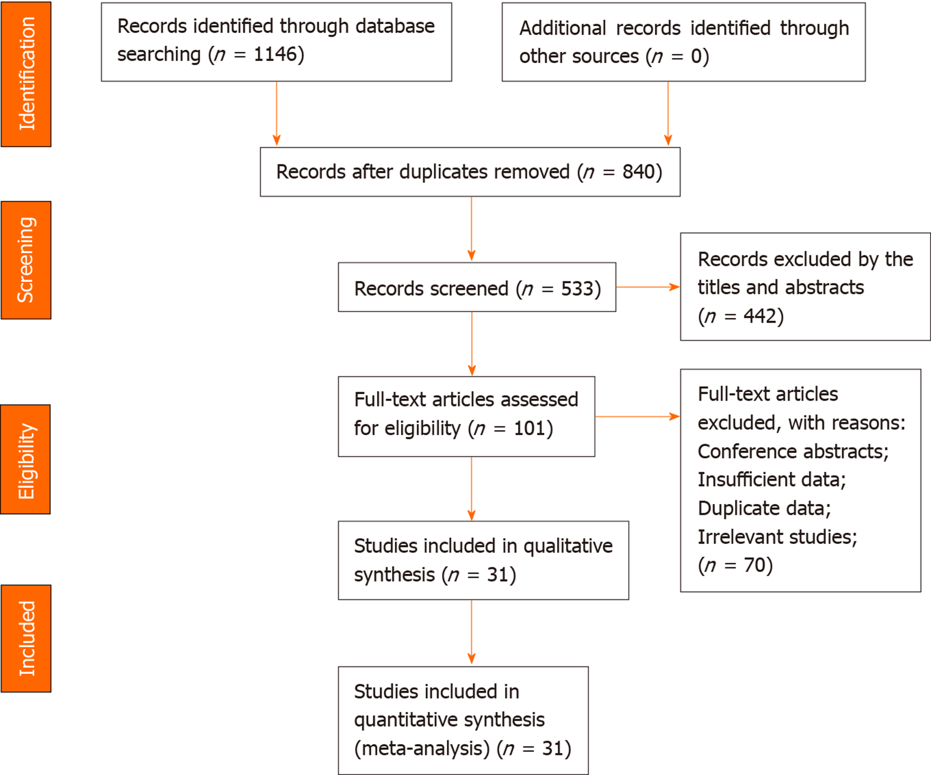
Figure 1 Study identification, inclusion and exclusion for meta-analysis.
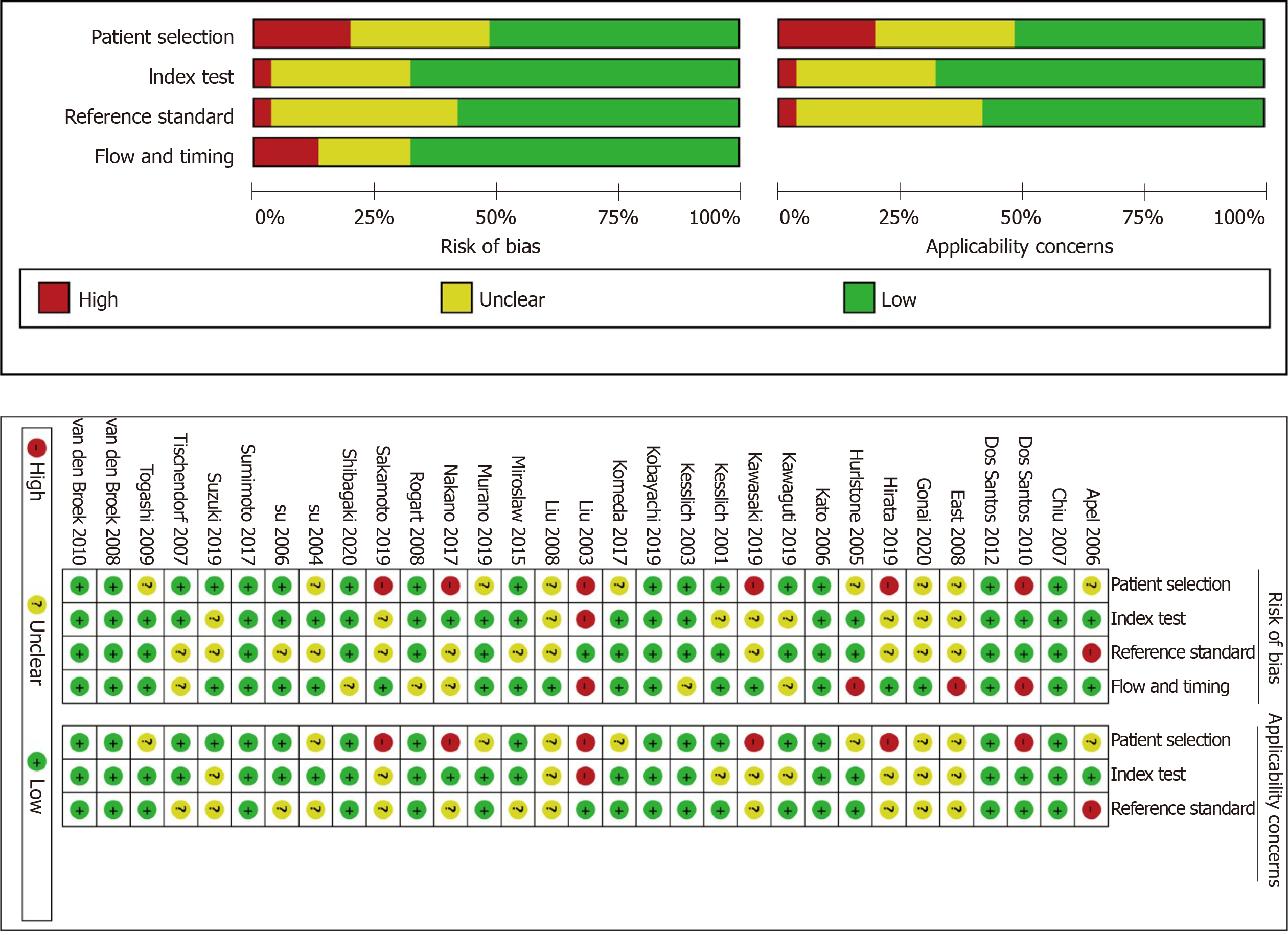
Figure 2 Quality assessment of the included studies.
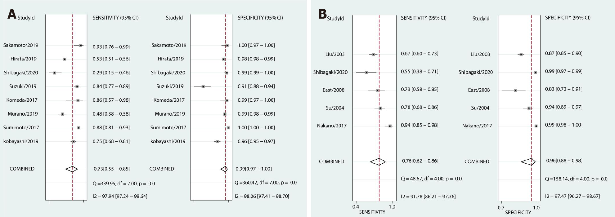
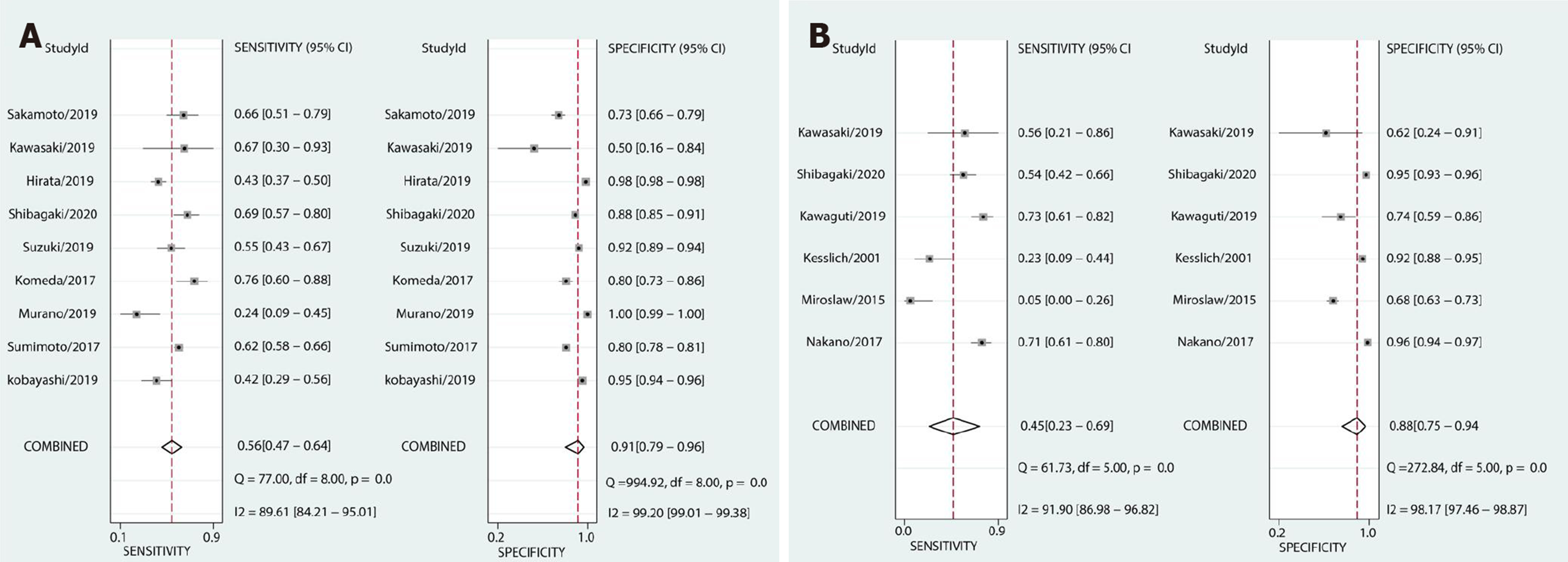
Figure 3 Forest plots of pooled sensitivity and specificity.
A: Japan Narrow-band-imaging Expert Team type 1; B: Pit pattern II.
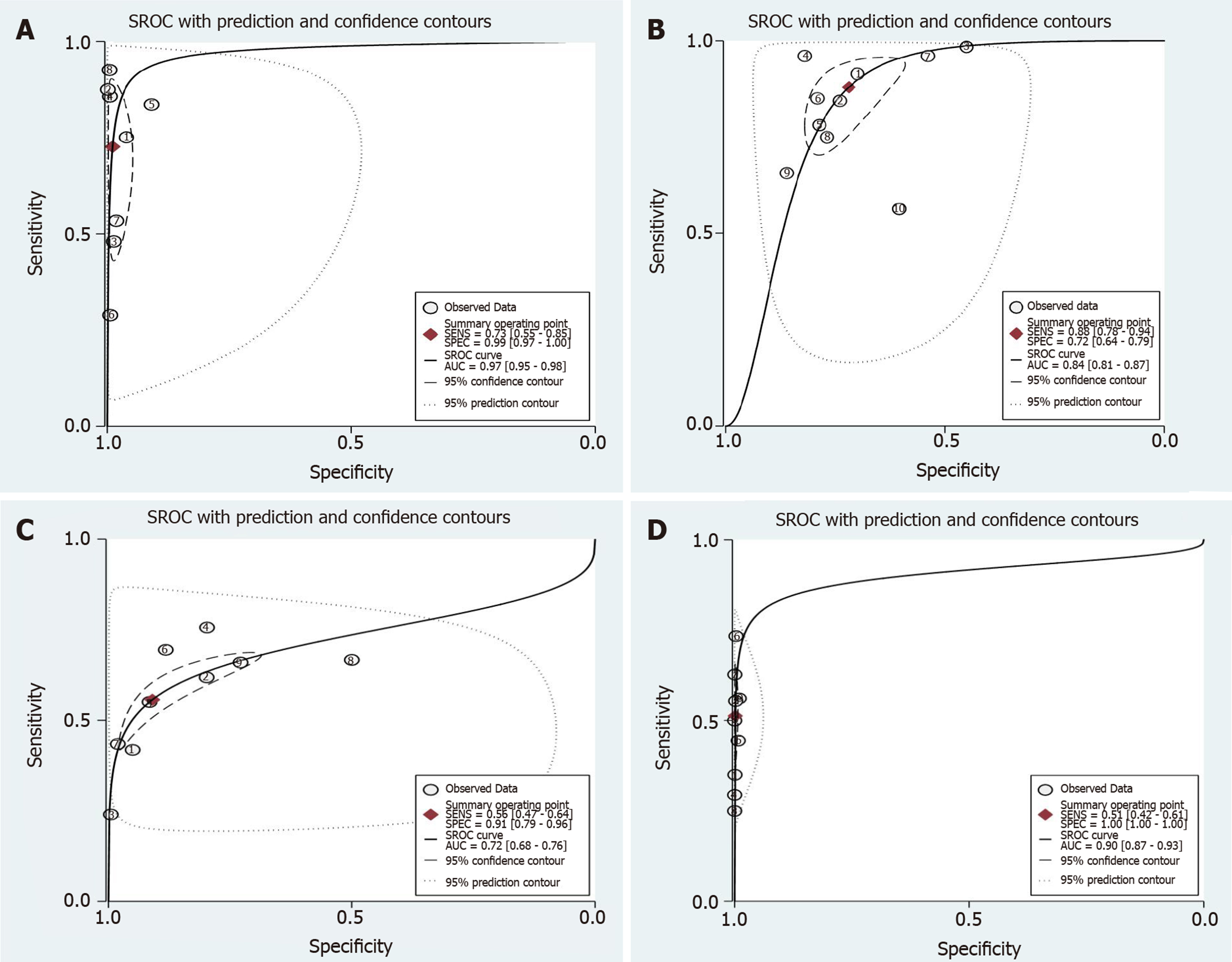
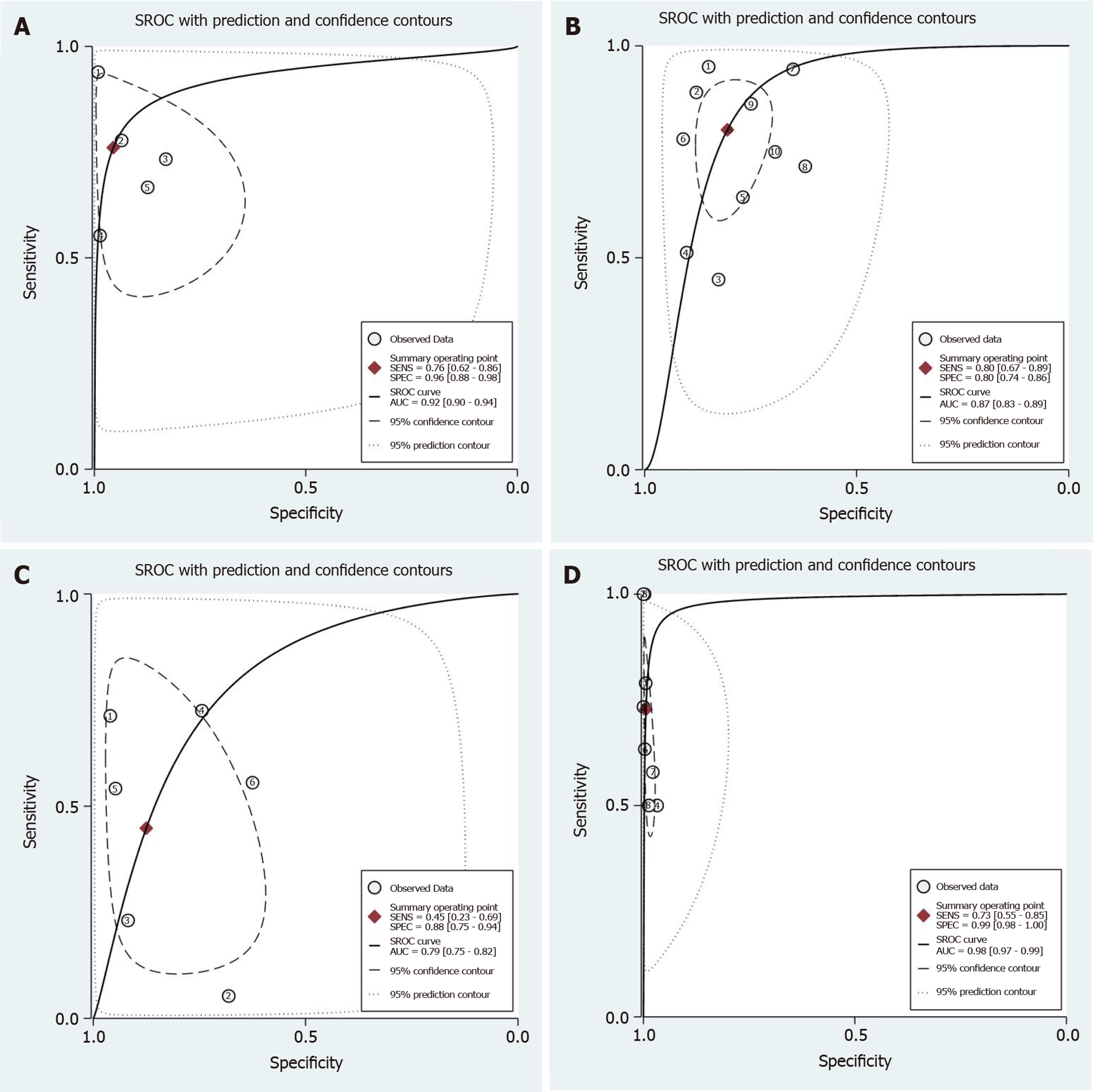
Figure 4 Summary receiver operating characteristic of the Japan Narrow-band-imaging Expert Team classification.
To diagnose colorectal lesions with the corresponding 95% confidence region. A: Type 1; B: Type 2A; C: Type 2B and Type 3. SROC: Summary receiver operating characteristic.
Figure 5 Forest plots of pooled sensitivity and specificity.
A: Japan Narrow-band-imaging Expert Team type 2B; B: Pit pattern IIIS + VI-L.
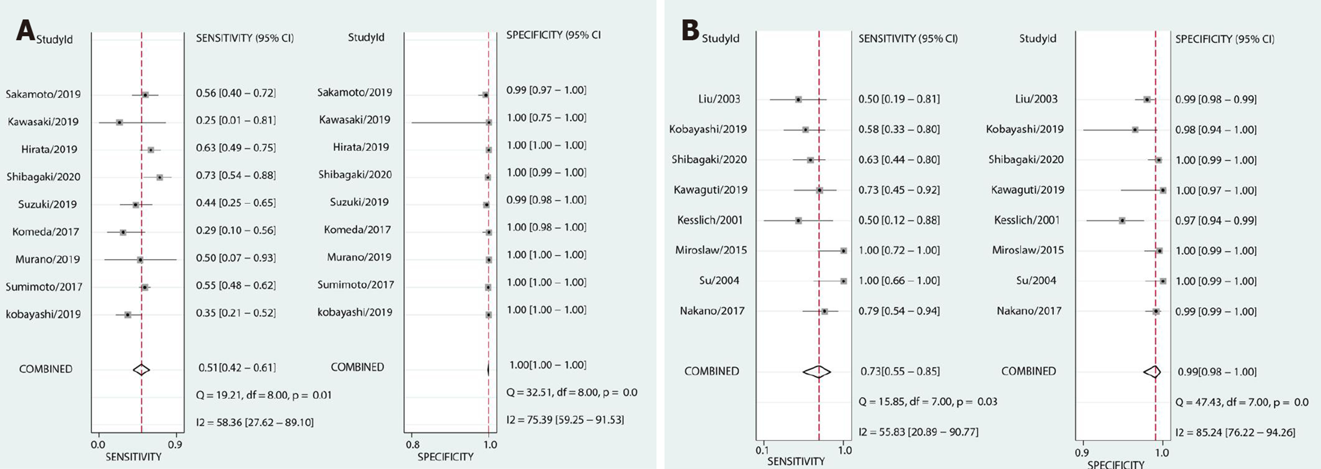
Figure 6 Forest plots of pooled sensitivity and specificity.
A: Japan Narrow-band-imaging Expert Team type 3; B: Pit pattern VN + VI-H.
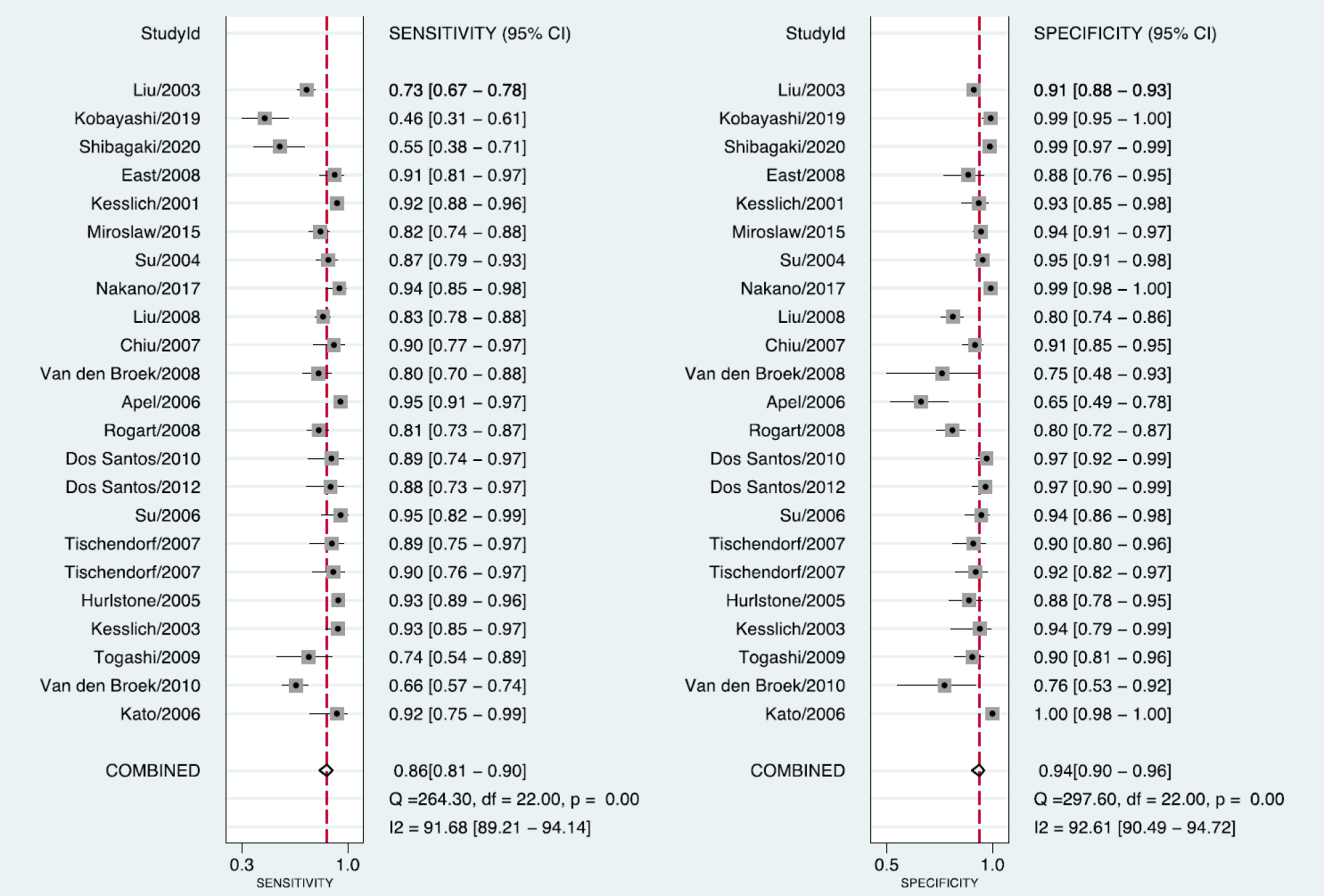
Figure 7 Forest plots of pooled sensitivity and specificity of non-neoplastic lesions by Pit pattern.
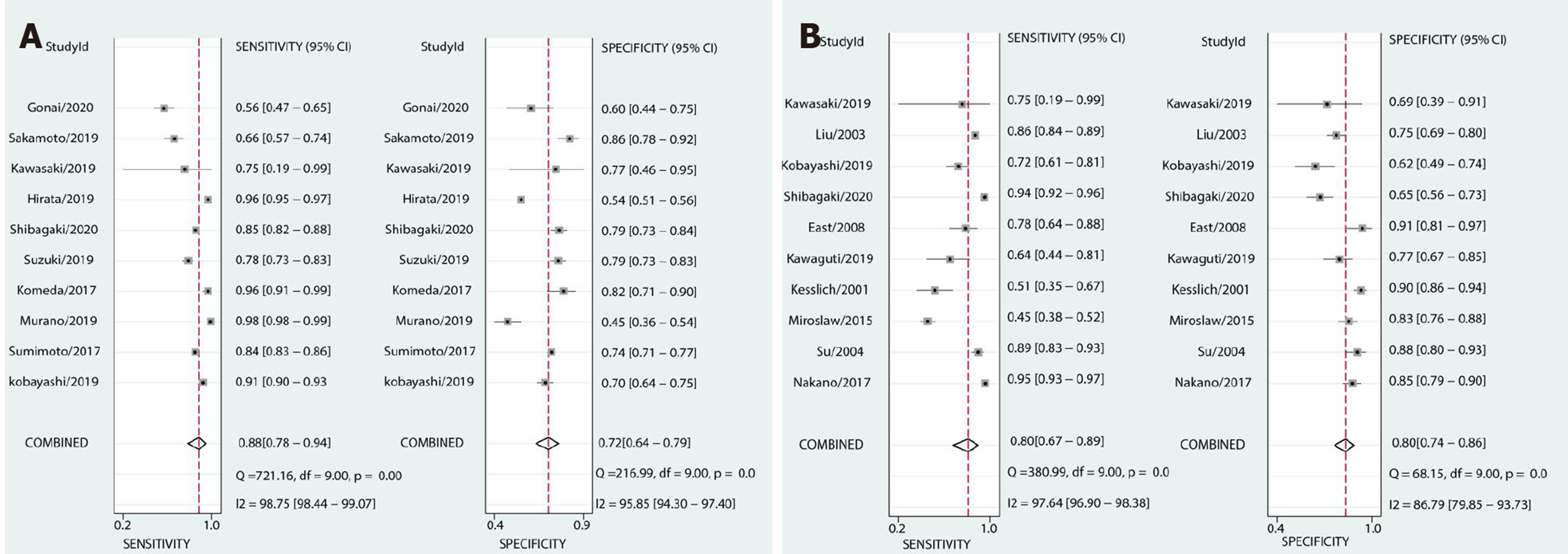
Figure 8 Forest plots of pooled sensitivity and specificity.
A: Japan Narrow-band-imaging Expert Team type 2A; B: Pit pattern IIIL + IV.
Figure 9 Summary receiver operating characteristic of Pit pattern classification.
To diagnose colorectal lesions with the corresponding 95% confidence region. A: II; B: IIIL + IV; C: IIIS + VI-L and VN + VI-H. SROC: Summary receiver operating characteristic.
- Citation: Zhang Y, Chen HY, Zhou XL, Pan WS, Zhou XX, Pan HH. Diagnostic efficacy of the Japan Narrow-band-imaging Expert Team and Pit pattern classifications for colorectal lesions: A meta-analysis. World J Gastroenterol 2020; 26(40): 6279-6294
- URL: https://www.wjgnet.com/1007-9327/full/v26/i40/6279.htm
- DOI: https://dx.doi.org/10.3748/wjg.v26.i40.6279


