Copyright
©The Author(s) 2020.
World J Gastroenterol. Oct 28, 2020; 26(40): 6241-6249
Published online Oct 28, 2020. doi: 10.3748/wjg.v26.i40.6241
Published online Oct 28, 2020. doi: 10.3748/wjg.v26.i40.6241
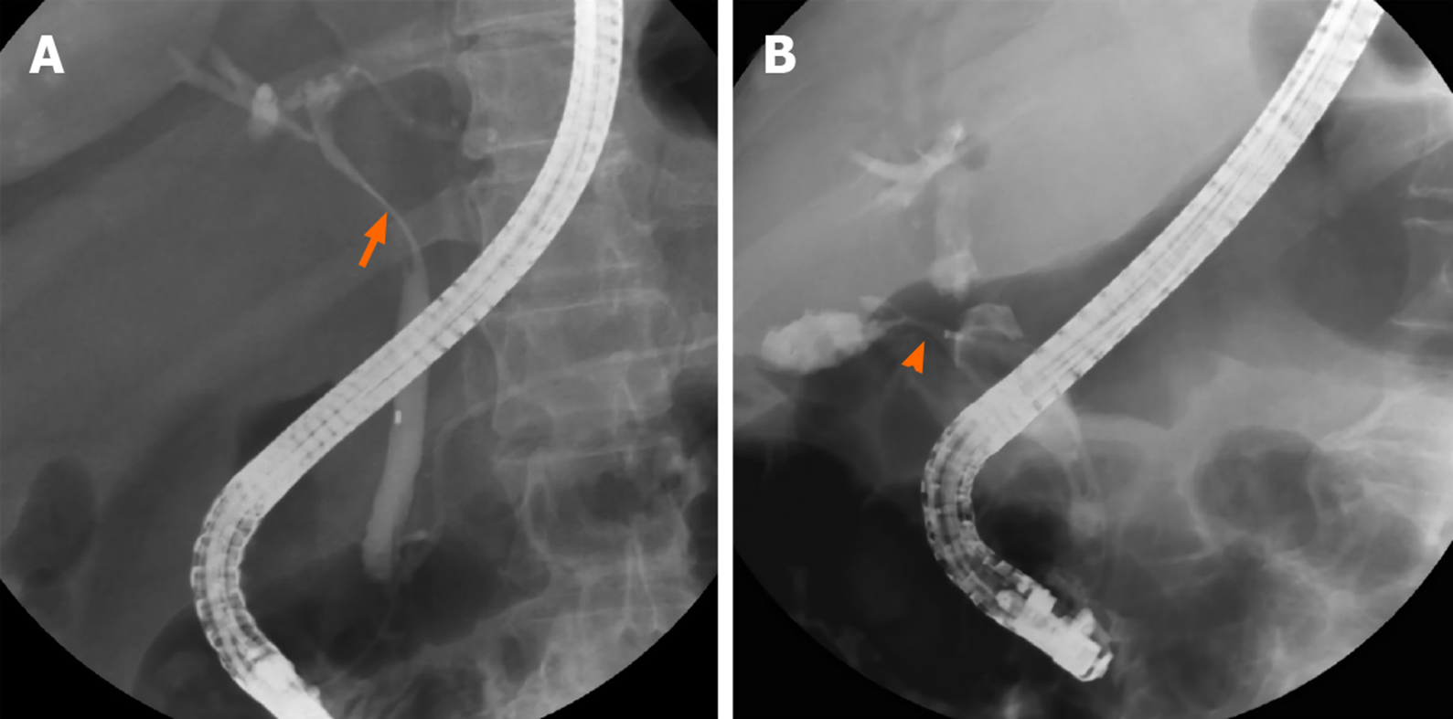
Figure 1 Cholangiography of patients with Mirizzi syndrome.
A: Patient without a cholecystocholedochal fistula. Eccentric compression (orange arrow) of the common bile duct is observed and a short part of the cystic duct is opacified; B: Patient with a cholecystocholedochal fistula (orange arrowhead). A contrast opacified gallbladder without the typical spiral and corkscrew-like cystic duct opacification is shown.
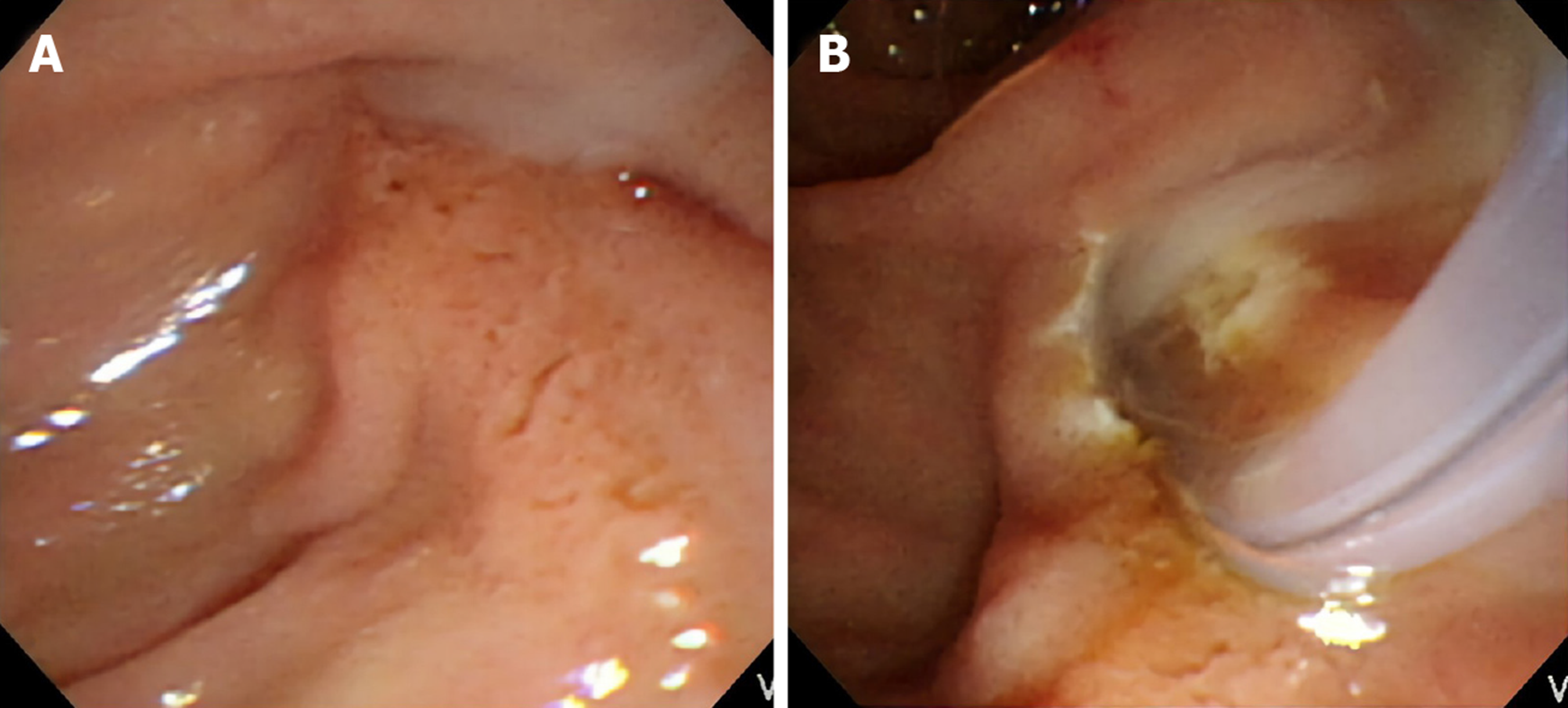
Figure 2 Endoscopic finding of pus in the common bile duct in patients with Mirizzi syndrome.
A: The pus is extruding from the ampulla of Vater during endoscopic retrograde cholangiopancreatography; B: The pus is observed from the ampulla of Vater after an endoscopic sphincterotomy.
Figure 3 Stricture length in common bile duct in patients with Mirizzi syndrome.
A: One 2.7-cm gall bladder stone caused a cholecystocholedochal fistula. The stricture length is only 1.7 cm, which is less than the actual size of the stone; B and C: One 1.4-cm gall bladder stone compressed the common bile duct. However, the stricture of common bile duct is 3.3 cm in length. Computerized tomography after endoscopic retrograde cholangiopancreatography revealed that the plastic biliary stent was close to the stone (orange arrow).
- Citation: Wu CH, Liu NJ, Yeh CN, Wang SY, Jan YY. Predicting cholecystocholedochal fistulas in patients with Mirizzi syndrome undergoing endoscopic retrograde cholangiopancreatography. World J Gastroenterol 2020; 26(40): 6241-6249
- URL: https://www.wjgnet.com/1007-9327/full/v26/i40/6241.htm
- DOI: https://dx.doi.org/10.3748/wjg.v26.i40.6241


