Copyright
©The Author(s) 2020.
World J Gastroenterol. Jan 28, 2020; 26(4): 433-447
Published online Jan 28, 2020. doi: 10.3748/wjg.v26.i4.433
Published online Jan 28, 2020. doi: 10.3748/wjg.v26.i4.433
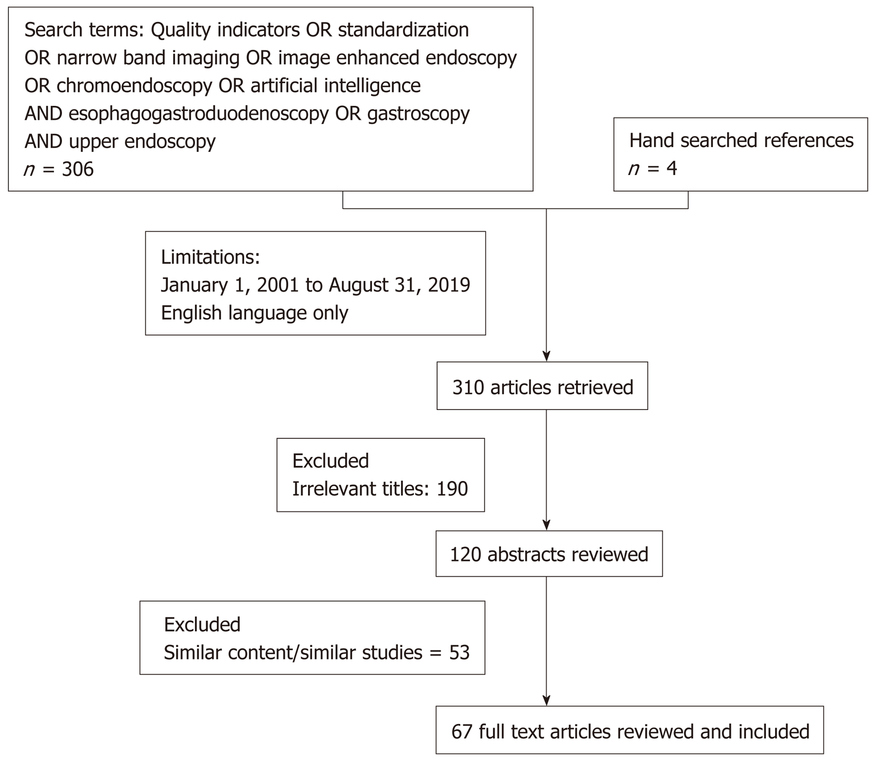
Figure 1 PRISMA diagram of literature review.
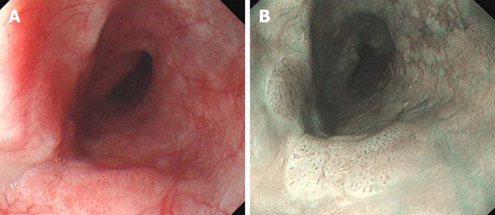
Figure 2 White light endoscopy and narrow band imaging of superficial esophageal squamous cell carcinoma.
A: White light endoscopy of superficial esophageal squamous cell carcinoma; B: Narrow band imaging of superficial esophageal squamous cell carcinoma.
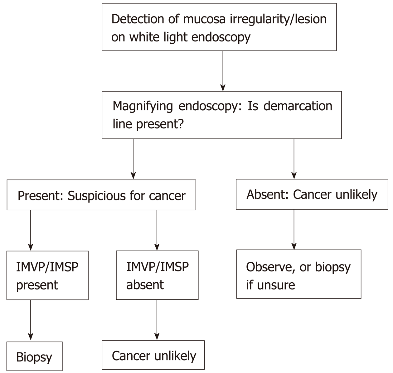
Figure 3 Magnifying endoscopy simple diagnostic algorithm for diagnosis of early gastric cancer.
Adapted from Muto et al[59]’s magnifying endoscopy simple diagnostic algorithm of early gastric cancer. IMVP: Irregular microvascular pattern; IMSP: Irregular microsurface pattern.
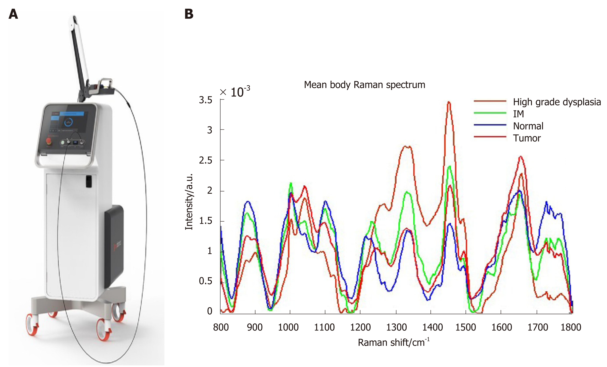
Figure 4 Raman spectroscopy probe and different Raman spectrum according to normal tissue, intestinal metaplasia, high grade dysplasia and tumor tissue.
A: Raman spectroscopy probe; B: Different Raman spectrum according to normal tissue, intestinal metaplasia, high grade dysplasia and tumor tissue.
- Citation: Teh JL, Shabbir A, Yuen S, So JBY. Recent advances in diagnostic upper endoscopy. World J Gastroenterol 2020; 26(4): 433-447
- URL: https://www.wjgnet.com/1007-9327/full/v26/i4/433.htm
- DOI: https://dx.doi.org/10.3748/wjg.v26.i4.433


