Copyright
©The Author(s) 2020.
World J Gastroenterol. Jan 28, 2020; 26(4): 424-432
Published online Jan 28, 2020. doi: 10.3748/wjg.v26.i4.424
Published online Jan 28, 2020. doi: 10.3748/wjg.v26.i4.424
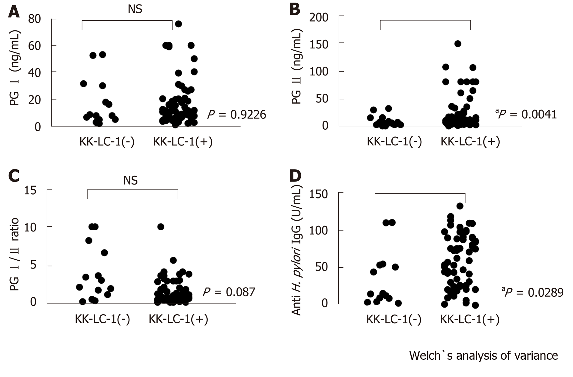
Figure 1 Variances of serum levels between the group with positive and negative Kita–Kyushu lung cancer antigen-1 expression in the gastric cancer patients.
The titers of pepsinogen (PG) I (A), PG II (B), the ratio of PG I/PG II (C), and anti-Helicobacter pylori antibody (IgG) (D) were measured in the Kita–Kyushu lung cancer antigen-1 (+) and Kita–Kyushu lung cancer antigen-1 (-) groups. aP < 0.05; NS: Not significant using the Welch’s t-test.
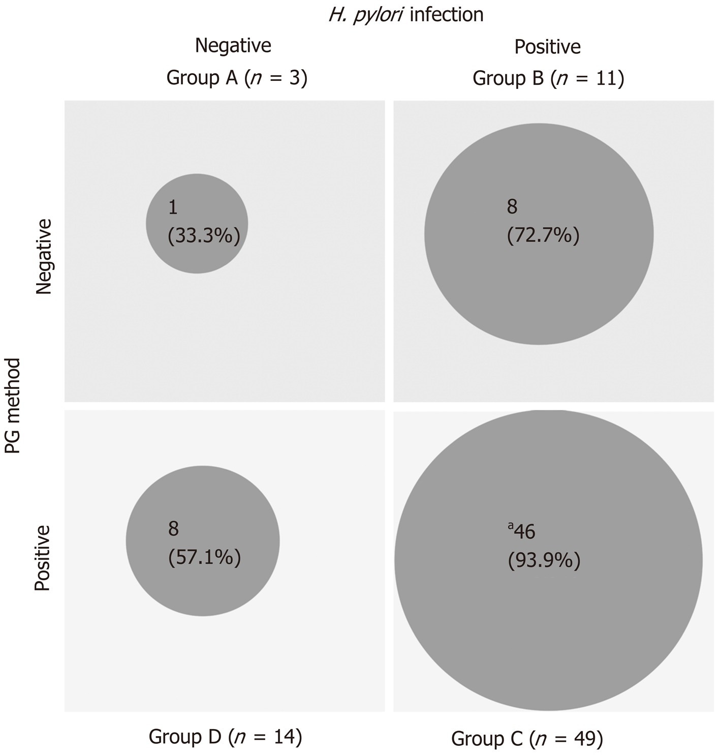
Figure 2 The frequency of Kita–Kyushu lung cancer antigen-1 expression according to the ABCD stratification.
The patients were classified into the A, B, C, and D groups using a combination of the status of Helicobacter pylori infection and PG value. The frequency of Kita–Kyushu lung cancer antigen-1 expression in each group was analyzed. aP = 0.0005, compared with the total study population (excluding group C) using the Fisher’s exact test. H. pylori: Helicobacter pylori.
- Citation: Shida A, Fukuyama T, Futawatari N, Ohmiya H, Ichiki Y, Yamashita T, Nishi Y, Kobayashi N, Yamazaki H, Watanabe M, Takahashi Y. Cancer/testis antigen, Kita-Kyushu lung cancer antigen-1 and ABCD stratification for diagnosing gastric cancers. World J Gastroenterol 2020; 26(4): 424-432
- URL: https://www.wjgnet.com/1007-9327/full/v26/i4/424.htm
- DOI: https://dx.doi.org/10.3748/wjg.v26.i4.424


