Copyright
©The Author(s) 2020.
World J Gastroenterol. Aug 7, 2020; 26(29): 4198-4217
Published online Aug 7, 2020. doi: 10.3748/wjg.v26.i29.4198
Published online Aug 7, 2020. doi: 10.3748/wjg.v26.i29.4198
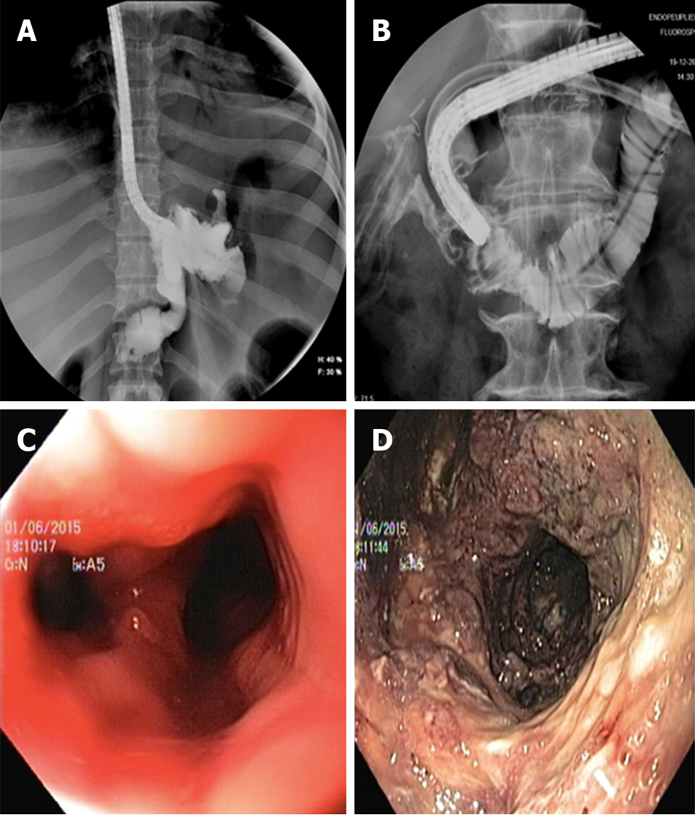
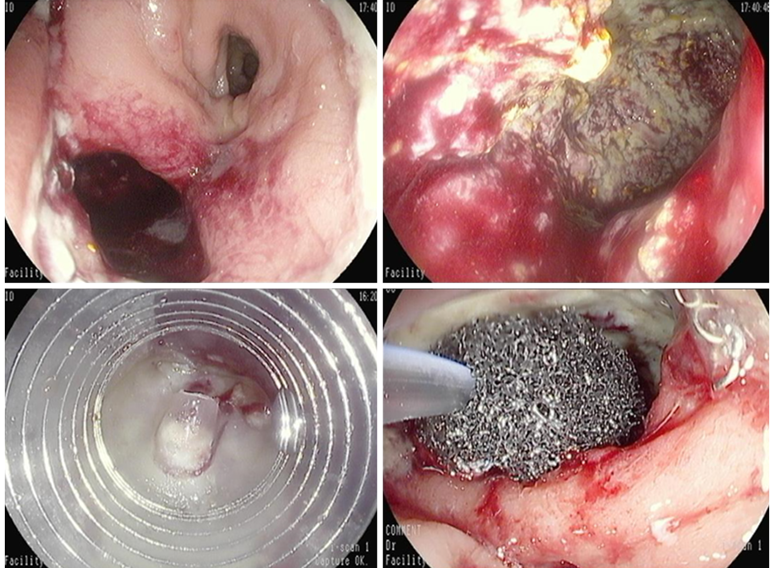
Figure 1 Radiological evidence and endoscopic view.
A: Radiological evidence of a gastric leak after sleeve gastrectomy; B: Radiological evidence of a duodenal leak after laparoscopic right hemicolectomy; C: Endoscopic view of leak orifice after sleeve gastrectomy; and D: Endoscopic exploration of leak associated collection.
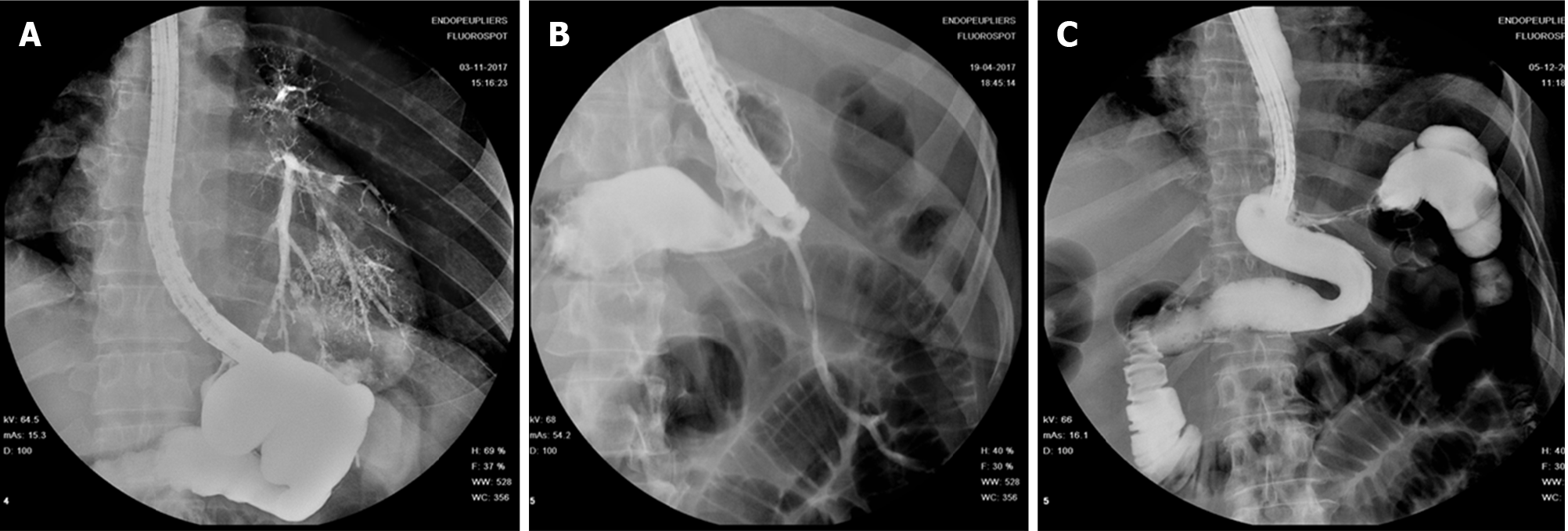
Figure 2 A fistula may involve many adjacent structures.
A: Gastro-bronchial fistula; B: Gastro-cutaneous fistula; and C: Gastro-colic fistula.
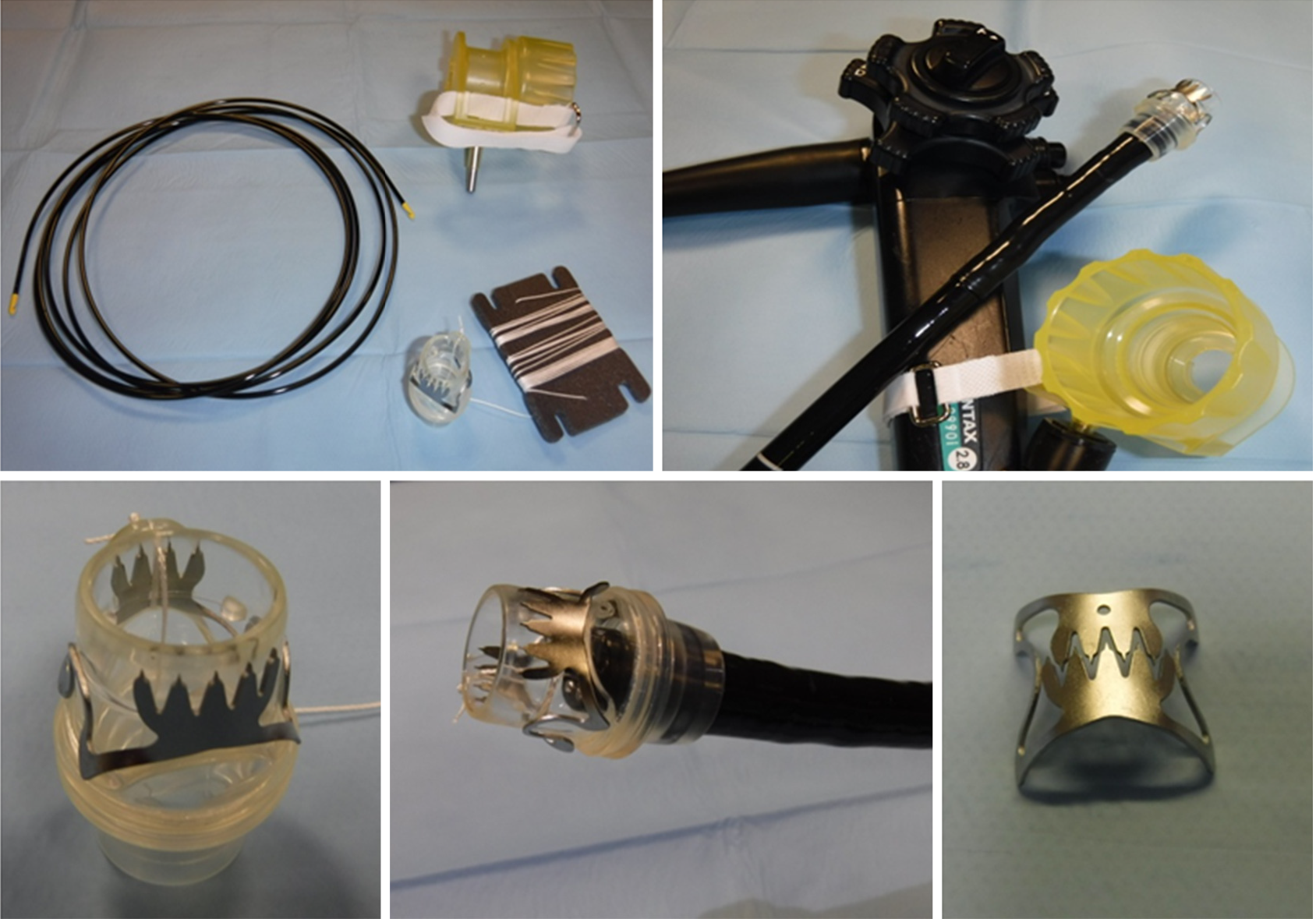
Figure 3 Over the scope clip system (Over-the-scope clips, Ovesco Endoscopy AG, Tubingen, Germany).
Figure 4 Over-the-scope clips closure of a leak after sleeve gastrectomy.
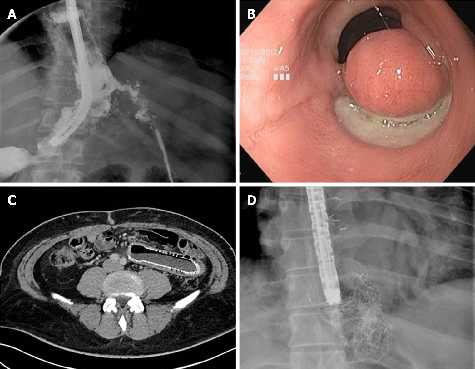
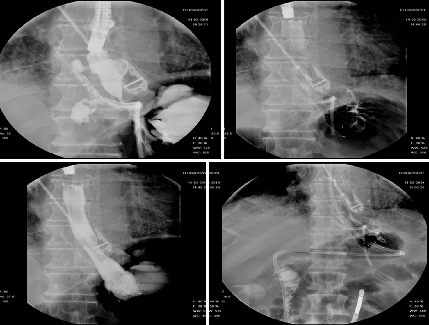
Figure 5 Self-expandable metal stent related adverse event.
A: Proximal stent migration with leak recurrence; B: Mucosal erosion and tissue overgrowth at the distal end of the stent after fully covered self-expandable metal stent removal; C: Distal stent migration and self-expandable metal stent related perforation; and D: Stent rupture during its removal.
Figure 6 Niti-S-Beta stent.
(Taewoong medical, Seoul, South Korea) deployment for the management of an early leak after sleeve gastrectomy.
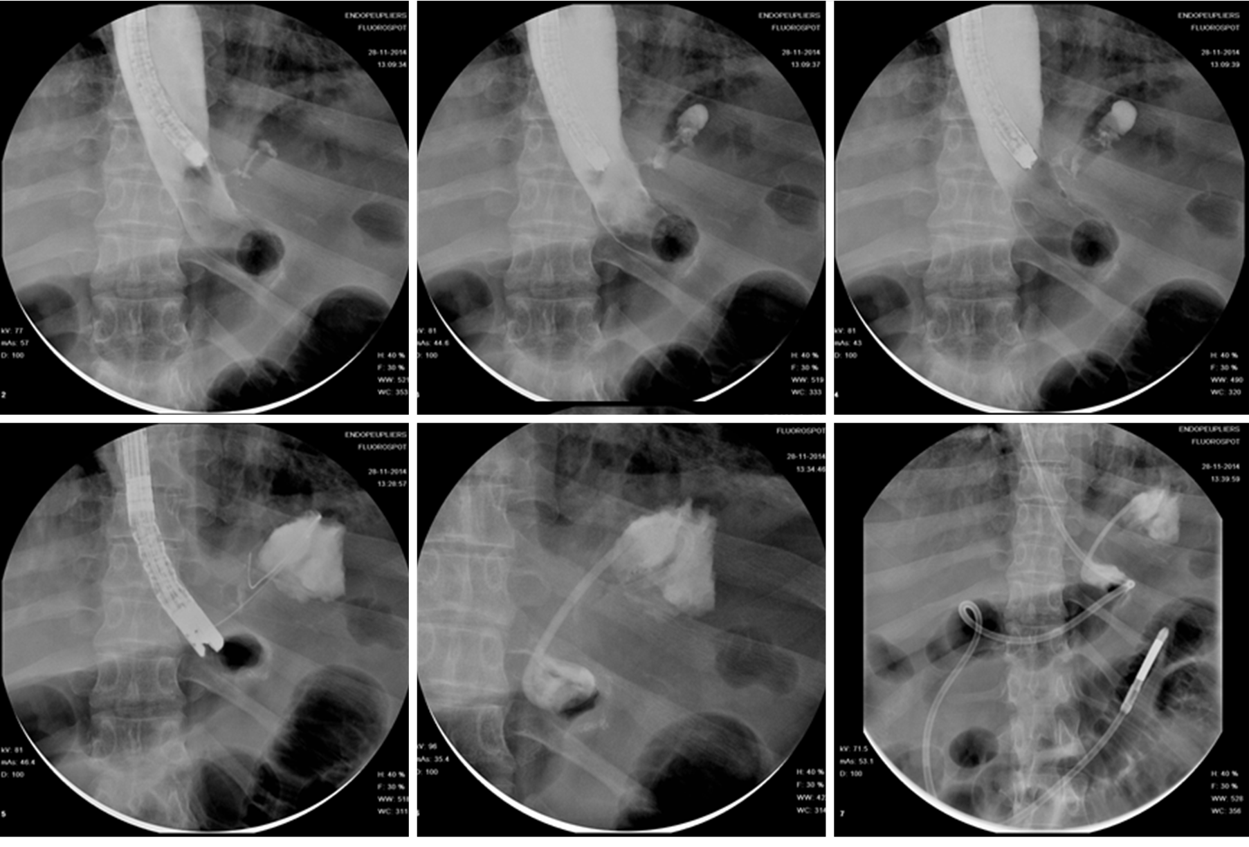
Figure 7 Endoscopic internal drainage coupled with enteral nutrition for the management of a late leak following sleeve gastrectomy.
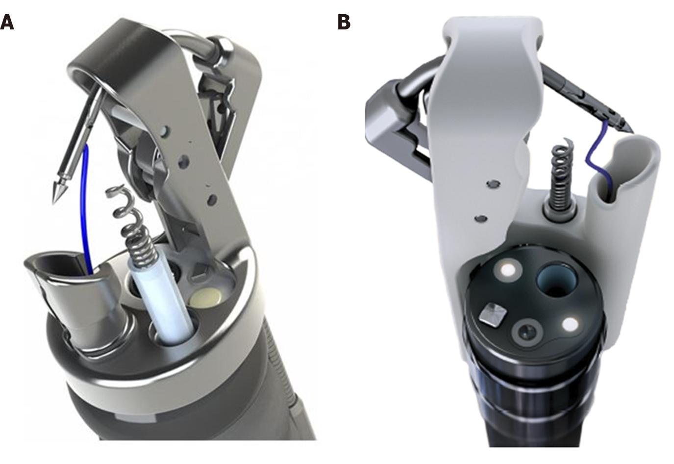
Figure 8 Overstich SX device.
A: OverStitch device (Apollo Endosurgery, Texas, United States); and B: Overstich SX device (Apollo Endosurgery, Texas, United States).
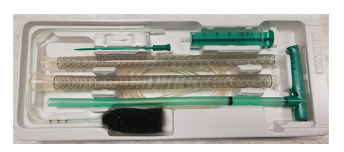
Figure 9 Endo-SPONGE® (B.
Braun Medical B.V., Melsungen, Germany).
Figure 10 Endoscopic vacuum assisted therapy for the management of an anastomotic leak after low anterior rectal resection.
- Citation: Cereatti F, Grassia R, Drago A, Conti CB, Donatelli G. Endoscopic management of gastrointestinal leaks and fistulae: What option do we have? World J Gastroenterol 2020; 26(29): 4198-4217
- URL: https://www.wjgnet.com/1007-9327/full/v26/i29/4198.htm
- DOI: https://dx.doi.org/10.3748/wjg.v26.i29.4198


