Copyright
©The Author(s) 2020.
World J Gastroenterol. Jun 28, 2020; 26(24): 3484-3494
Published online Jun 28, 2020. doi: 10.3748/wjg.v26.i24.3484
Published online Jun 28, 2020. doi: 10.3748/wjg.v26.i24.3484
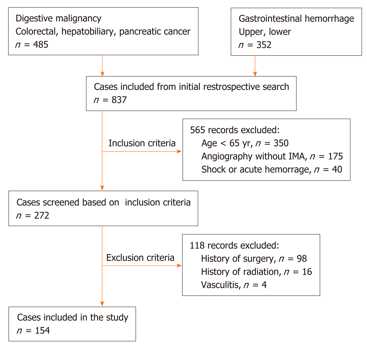
Figure 1 Flow chart summarizing patient enrollment.
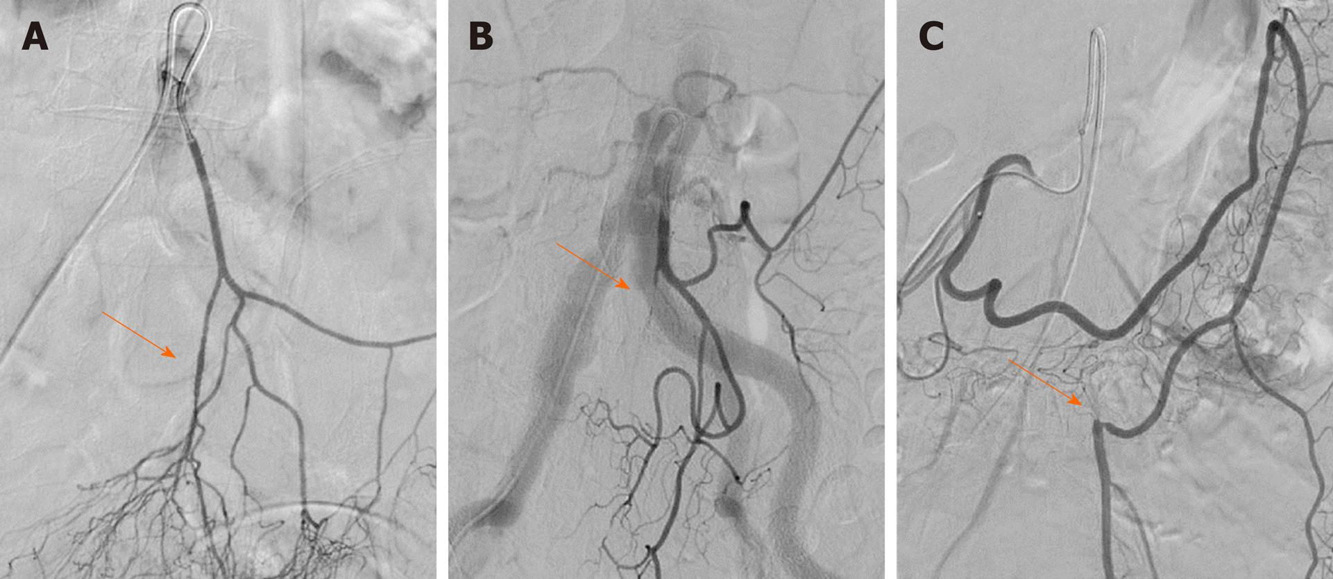
Figure 2 Demonstration of lesions in the inferior mesenteric artery and branches of the inferior mesenteric artery on digital subtraction angiography.
The arrows represent the lesion sites including stenosis and occlusion. A: Superior rectal artery stenosis with plaque; B: Superior rectal artery occlusion; C: Inferior mesenteric artery trunk occlusion. The distal blood supply came from left colic artery compensation for the superior mesenteric artery.
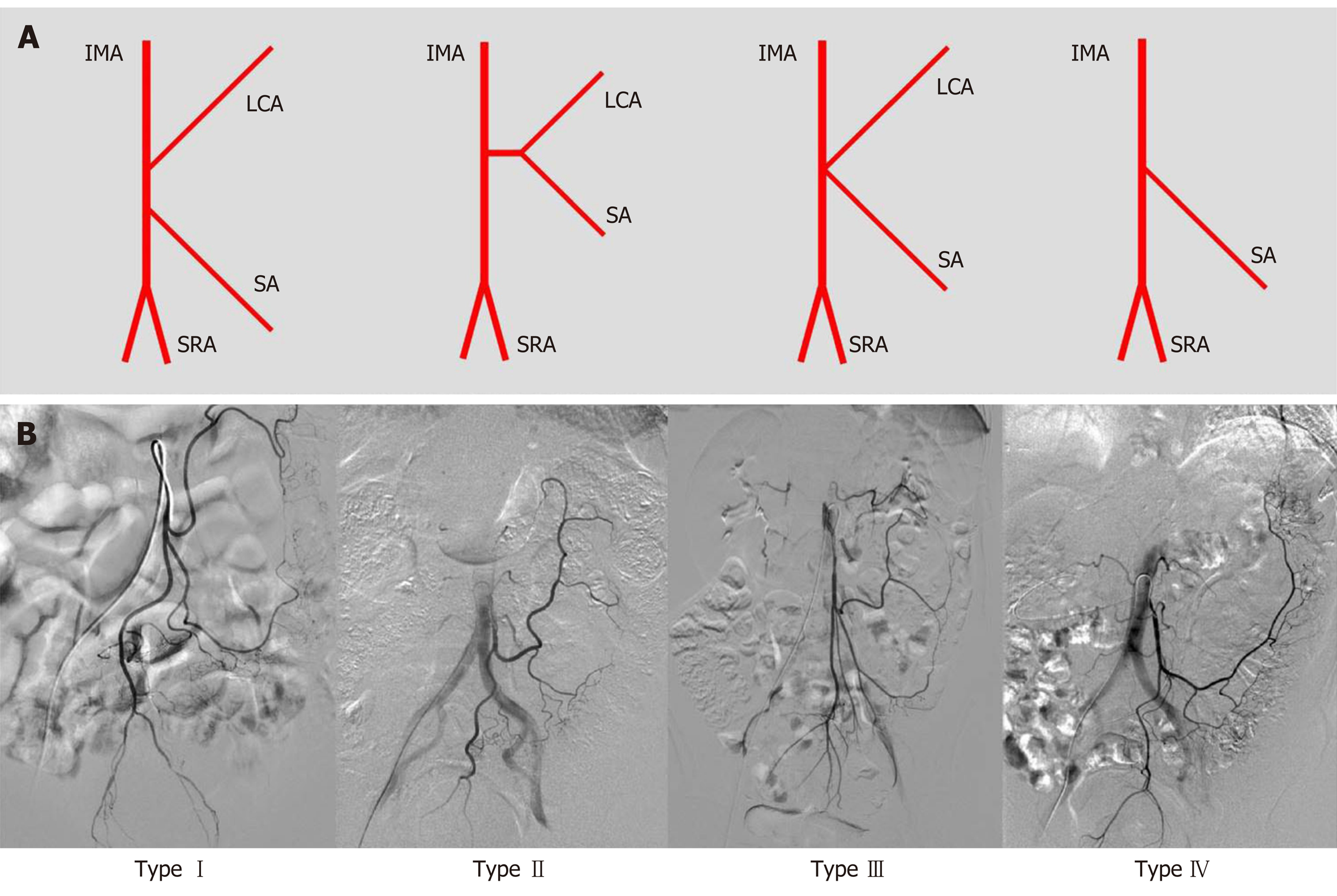
Figure 3 Patterns of inferior mesenteric artery bifurcation.
A: Illustration of the bifurcation of the inferior mesenteric artery (IMA) [only one sigmoid artery (SA) was showed in the illustration of IMA branches]. Type I: The LCA arose independently from IMA; type II: The LCA and SA arose from the IMA at the same point; type III: The LCA, SA, and SRA were branched from a common trunk from the IMA; and type IV: LCA was lacking; B: Angiography of the bifurcation of the IMA. IMA: Inferior mesenteric artery; SRA: Superior rectal artery; LCA: Left colic artery; SA: Sigmoid artery.
Figure 4 Demonstration of the colonic hemoperfusion region by angiography of the inferior mesenteric artery.
The arrows represent the inferior mesenteric artery (IMA) perfusion to the proximal region of the colon. A: IMA perfusion exceeded the splenic flexure and reached the transverse colon; B: IMA perfusion only achieved the splenic flexure and stopped; C: IMA perfusion reached only the descending colon with the left colic artery; D: IMA perfusion reached only the descending colon without the left colic artery.
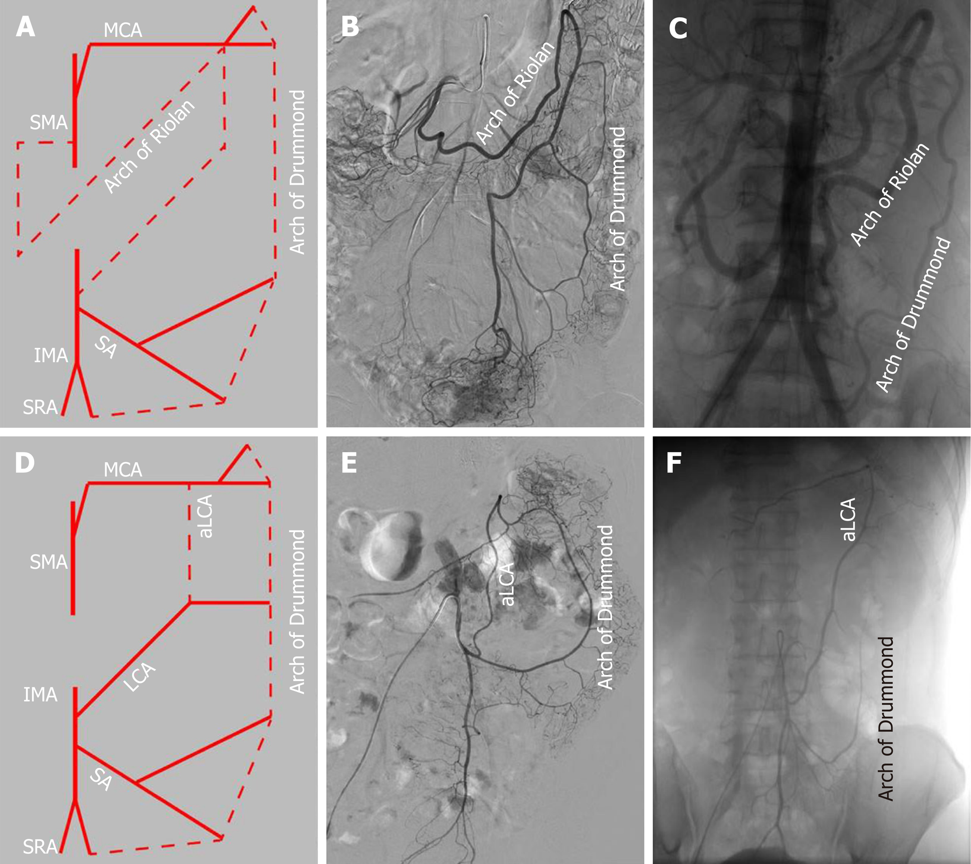
Figure 5 Patterns of the arterial arch connecting the superior mesenteric artery and inferior mesenteric artery, including the arc of Riolan (meandering mesenteric artery), Drummond’s artery (marginal artery), and ascending left colic artery.
A: Illustration of the arc of Riolan and Drummond’s artery; B: Angiography of IMA trunk occlusion with the arc of Riolan and Drummond’s artery; C: Angiography of SMA trunk occlusion with an abnormally enlarged arc of Riolan and Drummond’s artery; D: Illustration of the aLCA; E: Angiography of the normal Drummond artery with the aLCA to the transverse colon; F: Angiography of the aLCA with an incomplete Drummond artery. IMA: Inferior mesenteric artery; SMA: Superior mesenteric artery; SRA: Superior rectal artery; SA: Sigmoid artery; LCA: Left colic artery; aLCA: Ascending left colic artery; MCA: middle colic artery.
- Citation: Zhang C, Li A, Luo T, Li Y, Li F, Li J. Evaluation of characteristics of left-sided colorectal perfusion in elderly patients by angiography. World J Gastroenterol 2020; 26(24): 3484-3494
- URL: https://www.wjgnet.com/1007-9327/full/v26/i24/3484.htm
- DOI: https://dx.doi.org/10.3748/wjg.v26.i24.3484


