Copyright
©The Author(s) 2020.
World J Gastroenterol. May 28, 2020; 26(20): 2533-2549
Published online May 28, 2020. doi: 10.3748/wjg.v26.i20.2533
Published online May 28, 2020. doi: 10.3748/wjg.v26.i20.2533
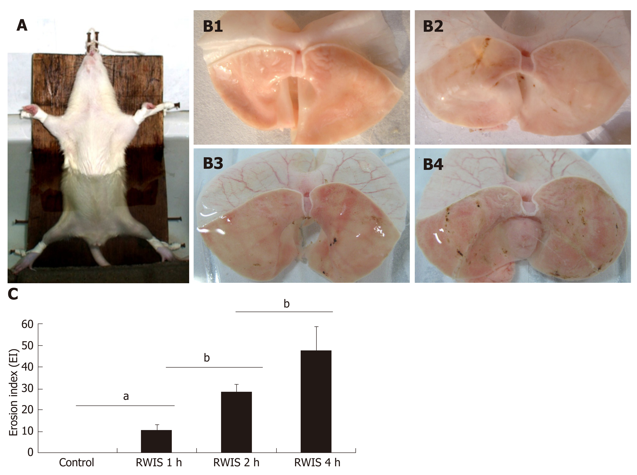
Figure 1 Effects of restraint water-immersion stress on gastric mucosal lesions in rats.
A: The animal model of restraint water-immersion stress (RWIS). B: Gastric mucosal damage induced by RWIS (B1: control; B2: 1-h RWIS; B3: 2-h RWIS; B4: 4-h RWIS). C: Effects of RWIS on the rates of gastric erosion. n = 5; mean ± SE; aP < 0.05, bP < 0.01. RWIS: Restraint water-immersion stress.
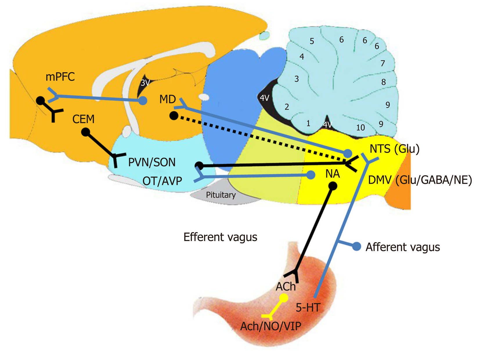
Figure 2 Schematic diagram of the central nervous pathways of restraint water-immersion stress-regulated gastrointestinal function.
Solid line: The pathways that have been determined; Dashed line: The pathways that remain to be further verified; Blue line segment: Upward; Green line segment: Downward. mPFC: Medial prefrontal cortex; CEA: Central nucleus of the amygdala; MD: Mediodorsal nucleus of the thalamus; DMV: Dorsal motor nucleus of vagus; NA: Nucleus ambiguous; NTS: Nucleus of the solitary tract; VIP: Vasoactive intestinal peptide; 5-HT: 5-hydroxytryptamine; NO: Nitric oxide; ACh: Acetylcholine; NE: Norepinephrine; GABA: γ-aminiobutyric acid; Glu: Glutamate; PVN: Paraventricular nucleus; SON: Supraoptic nucleus; OT: Oxytocin; AVP: Arginine vasopressin.
- Citation: Zhao DQ, Xue H, Sun HJ. Nervous mechanisms of restraint water-immersion stress-induced gastric mucosal lesion. World J Gastroenterol 2020; 26(20): 2533-2549
- URL: https://www.wjgnet.com/1007-9327/full/v26/i20/2533.htm
- DOI: https://dx.doi.org/10.3748/wjg.v26.i20.2533


