Copyright
©The Author(s) 2020.
World J Gastroenterol. Apr 28, 2020; 26(16): 1971-1978
Published online Apr 28, 2020. doi: 10.3748/wjg.v26.i16.1971
Published online Apr 28, 2020. doi: 10.3748/wjg.v26.i16.1971
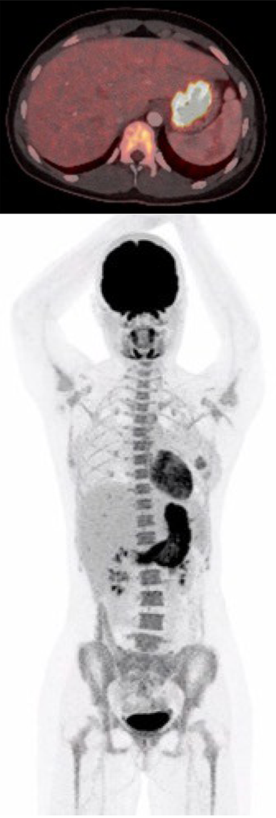
Figure 1 The first positron emission tomography with computed tomography after the patient presented with upper gastrointestinal symptoms.
The scan showed abnormal fluorodeoxyglucose uptake in the gastric wall, especially around the corpus antrum.
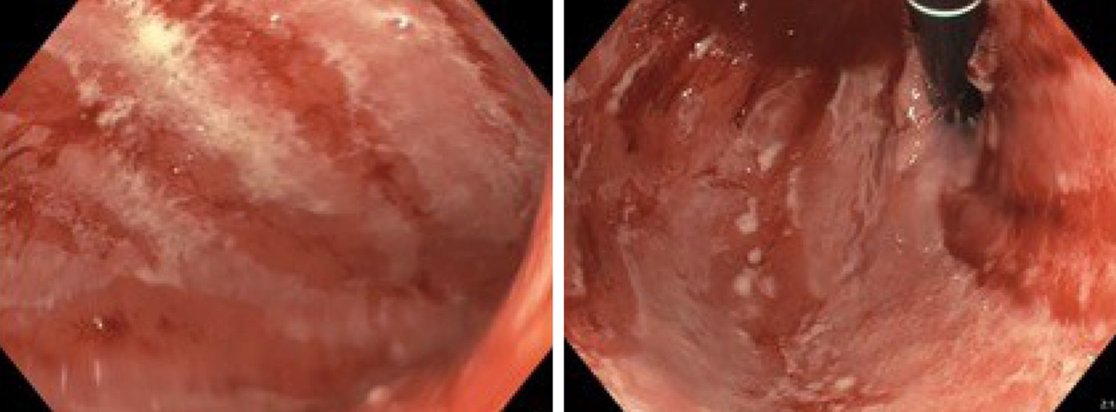
Figure 2 Esophagogastroduodenoscopy before treatment.
The gastric wall was erythematous with severe fibrinous erosions of the mucosa.
Figure 3 Imaging of histopathology.
A, B: Diffuse chronic active pangastritis with ulceration and only scattered glands. Neutrophilic inflammation and crypt abscesses increased intraepithelial lymphocytes and apoptosis (arrow) (A: 100 ×; B: 400 ×); C: Regenerated epithelium with focal acute inflammation (100 ×).
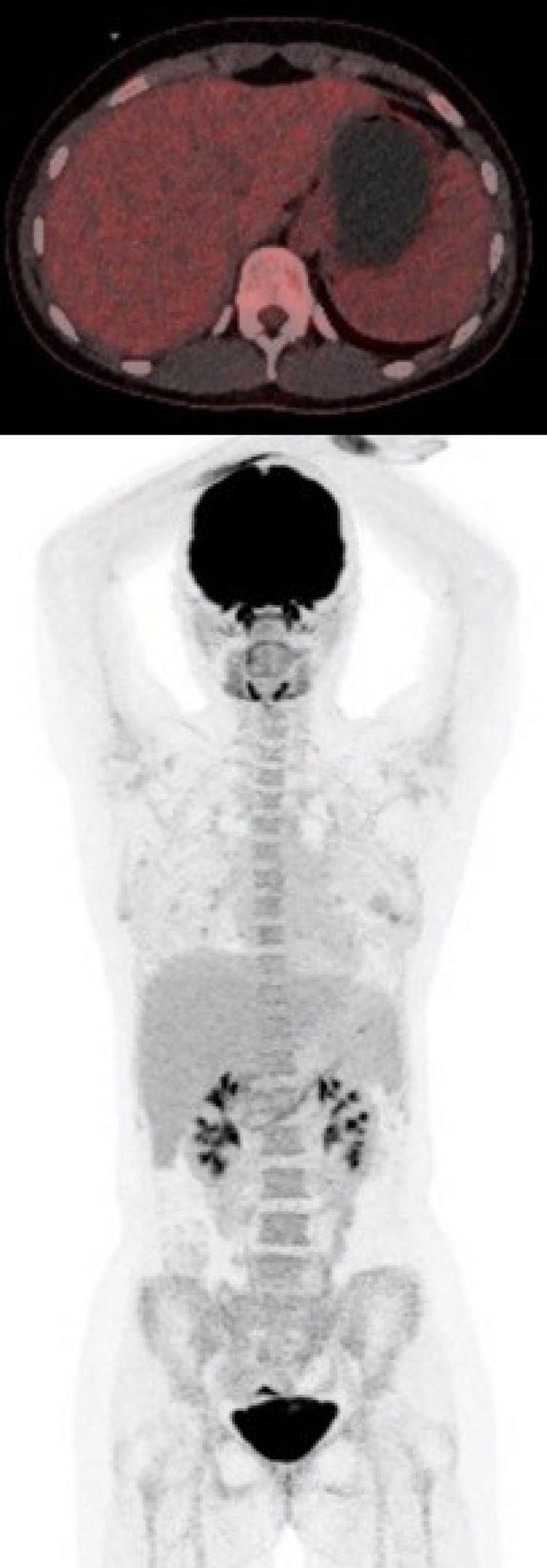
Figure 4 The second positron emission tomography with computed tomography performed 10 wk after the first Infliximab administration.
This showed a normal gastric wall with no fluorodeoxyglucose uptake.
- Citation: Vindum HH, Agnholt JS, Nielsen AWM, Nielsen MB, Schmidt H. Severe steroid refractory gastritis induced by Nivolumab: A case report. World J Gastroenterol 2020; 26(16): 1971-1978
- URL: https://www.wjgnet.com/1007-9327/full/v26/i16/1971.htm
- DOI: https://dx.doi.org/10.3748/wjg.v26.i16.1971


