Copyright
©The Author(s) 2020.
World J Gastroenterol. Apr 7, 2020; 26(13): 1540-1545
Published online Apr 7, 2020. doi: 10.3748/wjg.v26.i13.1540
Published online Apr 7, 2020. doi: 10.3748/wjg.v26.i13.1540
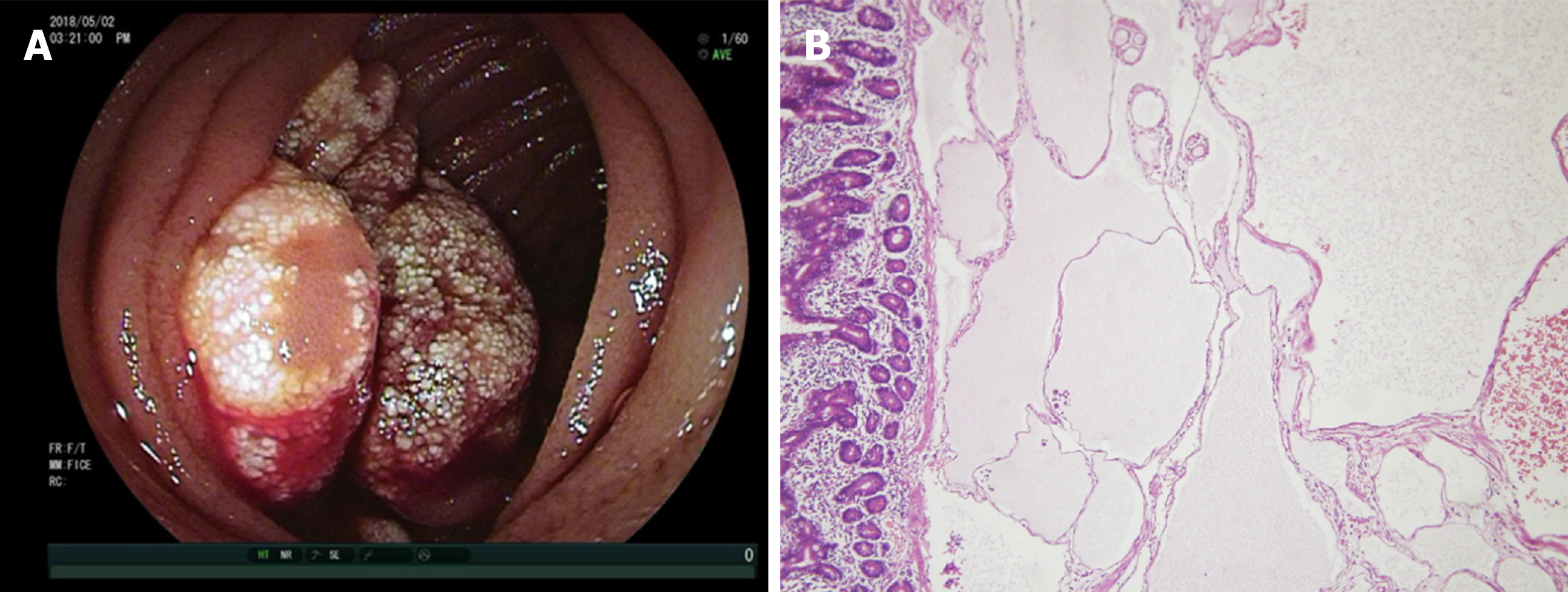
Figure 1 Gross and histologic images of hemolymphangioma.
A: Lobulated tumor occupied half of the intestinal cavity with white patches on the mucosal surface and blood oozing in the fundus; B: Histology revealed a hyperplastic thin-walled lymphangion and venous with luminal dilation in the submucosal area. Hematoxylin and eosin × 20.
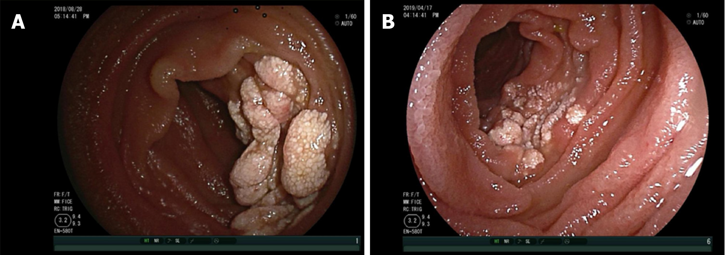
Figure 2 Images at the 3 mo and 1 year follow-up appointments.
A: At 3 mo after enteroscopic injection sclerotherapy, hemolymphangioma atrophied dramatically, and bleeding was hardly observed; B: At 1 year later, the hemolymphangioma was gone. A few white patches on the mucosal surface are visible.
- Citation: Xiao NJ, Ning SB, Li T, Li BR, Sun T. Small intestinal hemolymphangioma treated with enteroscopic injection sclerotherapy: A case report and review of literature. World J Gastroenterol 2020; 26(13): 1540-1545
- URL: https://www.wjgnet.com/1007-9327/full/v26/i13/1540.htm
- DOI: https://dx.doi.org/10.3748/wjg.v26.i13.1540


