Copyright
©The Author(s) 2019.
World J Gastroenterol. Mar 7, 2019; 25(9): 1088-1099
Published online Mar 7, 2019. doi: 10.3748/wjg.v25.i9.1088
Published online Mar 7, 2019. doi: 10.3748/wjg.v25.i9.1088
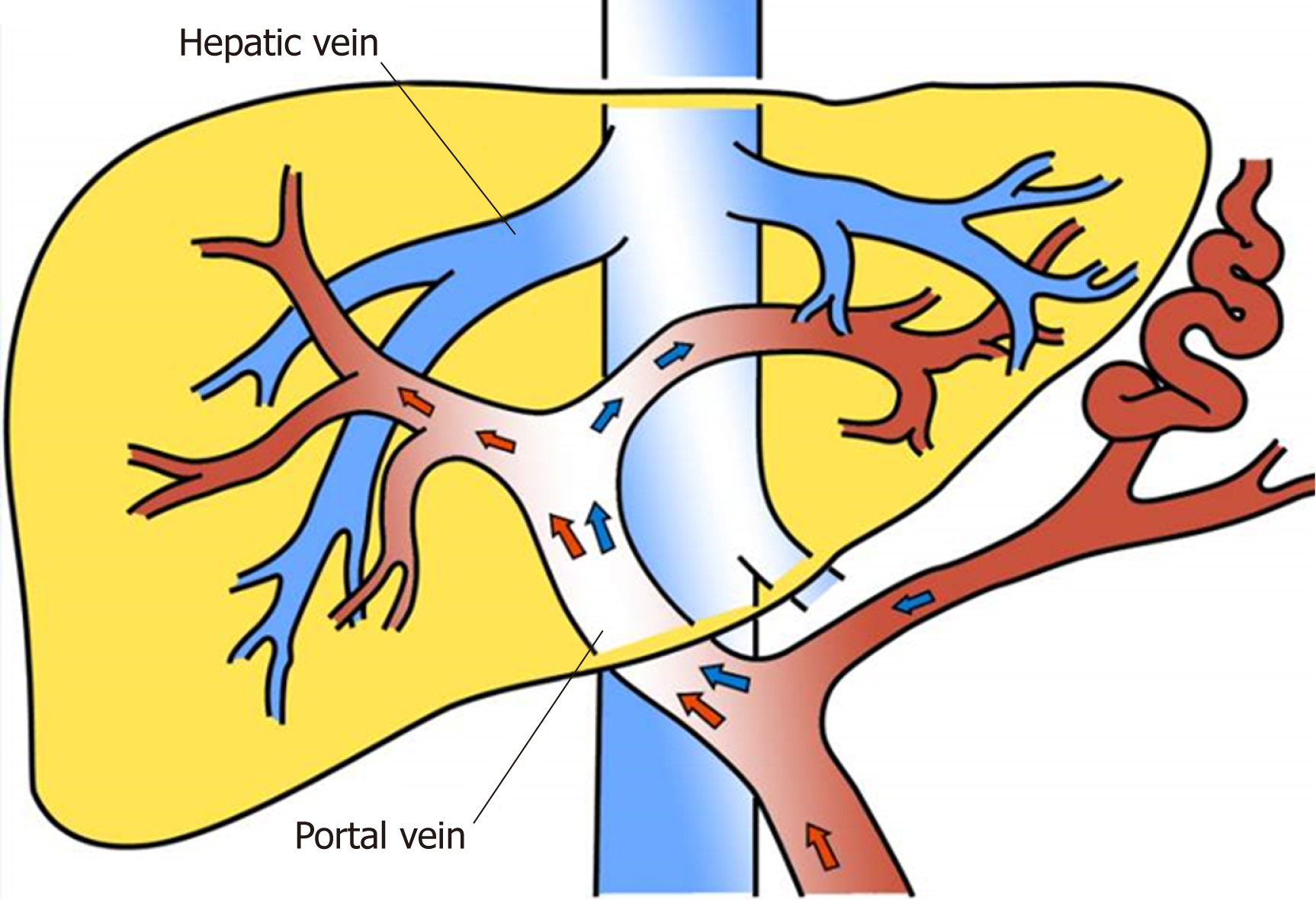
Figure 1 Blood distributed hydrodynamically in the main portal vein.
It is reported that the reflux blood from the splenic and superior mesenteric veins is distributed hydrodynamically in the main portal vein, that is, alongside the trunk on both sides of the wall of the portal vein. However, it is not fully mixed and enters the left and right branches of the portal vein. The right branch mainly receives superior mesenteric venous blood, while the left branch mainly receives blood from the splenic vein.
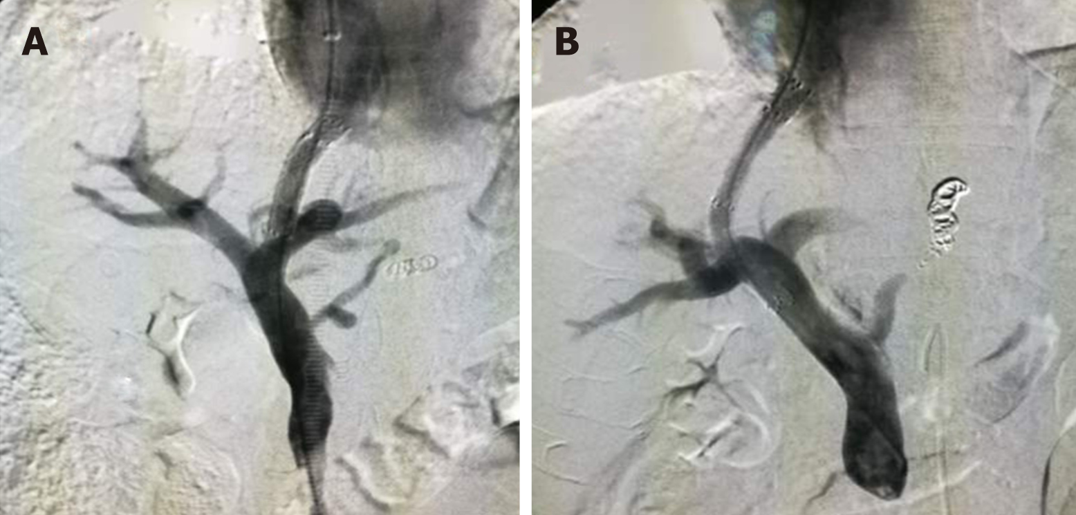
Figure 2 Shunt in the left or right branch of portal vein.
A: Shunt in the left branch of portal vein; B: Shunt in the right branch of portal vein.
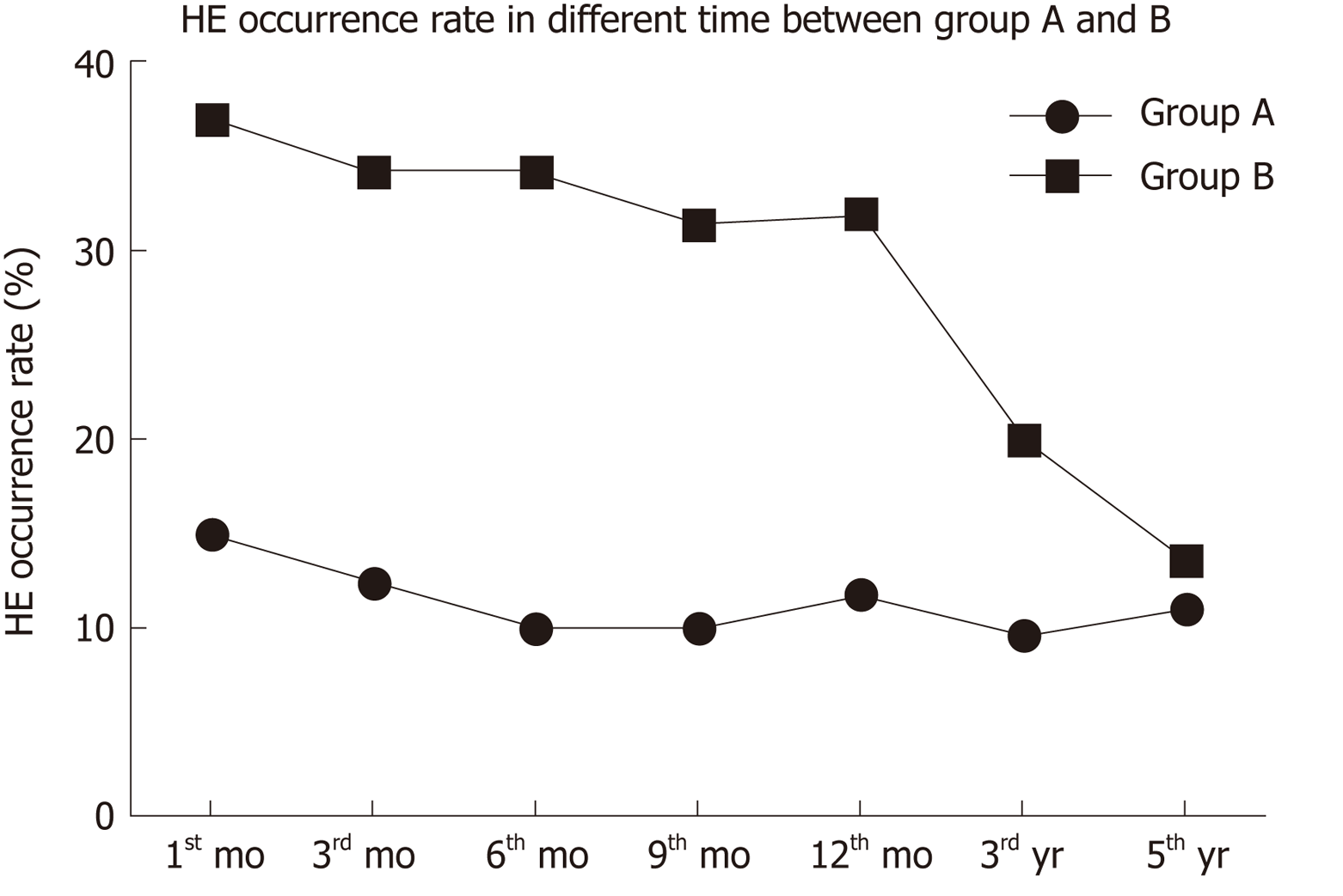
Figure 3 HE occurrence in the two groups.
HE occurrence rate in group A was lower than that in group B at 1, 3, 6, 9, and 12 mo, and the occurrence of HE showed a downward trend. HE: Hepatic encephalopathy.
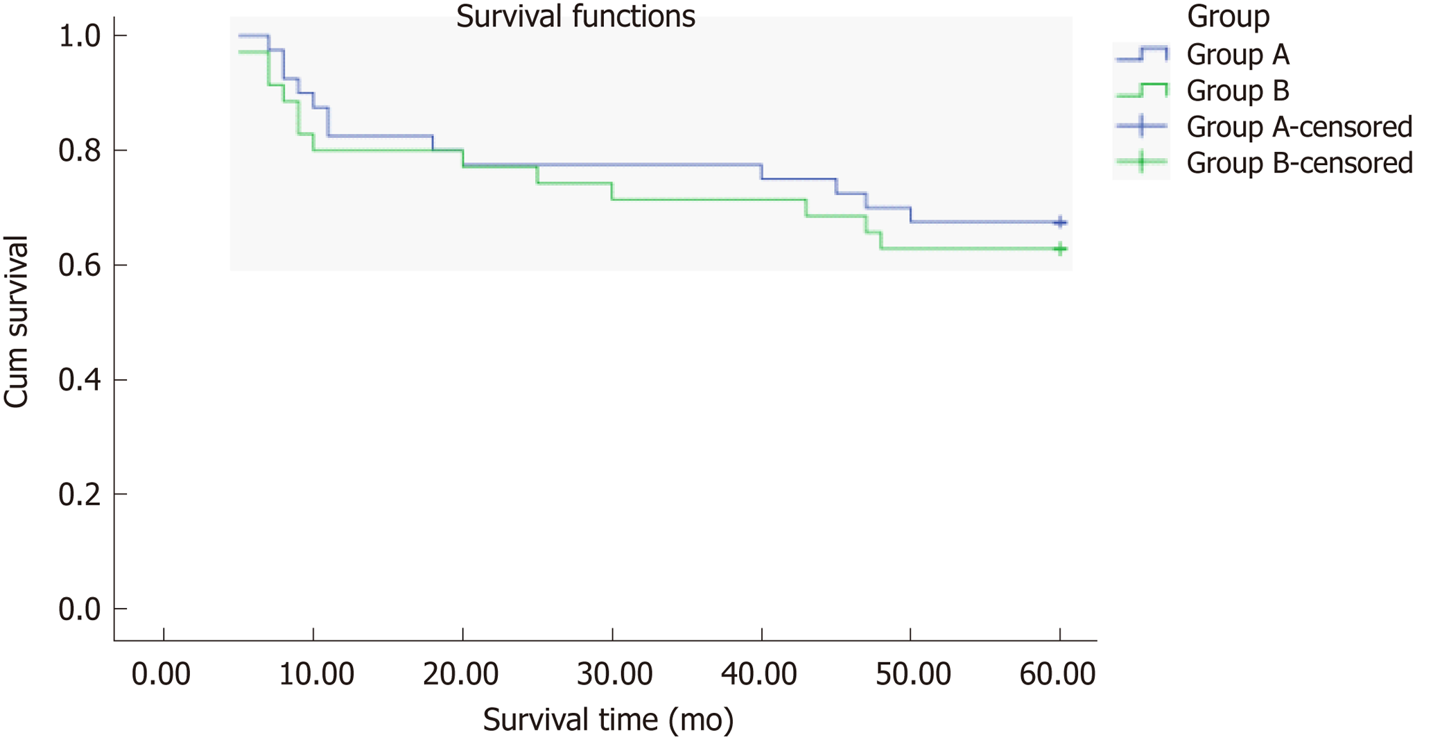
Figure 4 Total survival rate in the two groups.
The total survival rate did not differ between groups A and B (χ2 = 0.226, P = 0.634, log-rank test).
- Citation: Luo SH, Chu JG, Huang H, Zhao GR, Yao KC. Targeted puncture of left branch of intrahepatic portal vein in transjugular intrahepatic portosystemic shunt to reduce hepatic encephalopathy. World J Gastroenterol 2019; 25(9): 1088-1099
- URL: https://www.wjgnet.com/1007-9327/full/v25/i9/1088.htm
- DOI: https://dx.doi.org/10.3748/wjg.v25.i9.1088


