Copyright
©The Author(s) 2019.
World J Gastroenterol. Dec 7, 2019; 25(45): 6693-6703
Published online Dec 7, 2019. doi: 10.3748/wjg.v25.i45.6693
Published online Dec 7, 2019. doi: 10.3748/wjg.v25.i45.6693
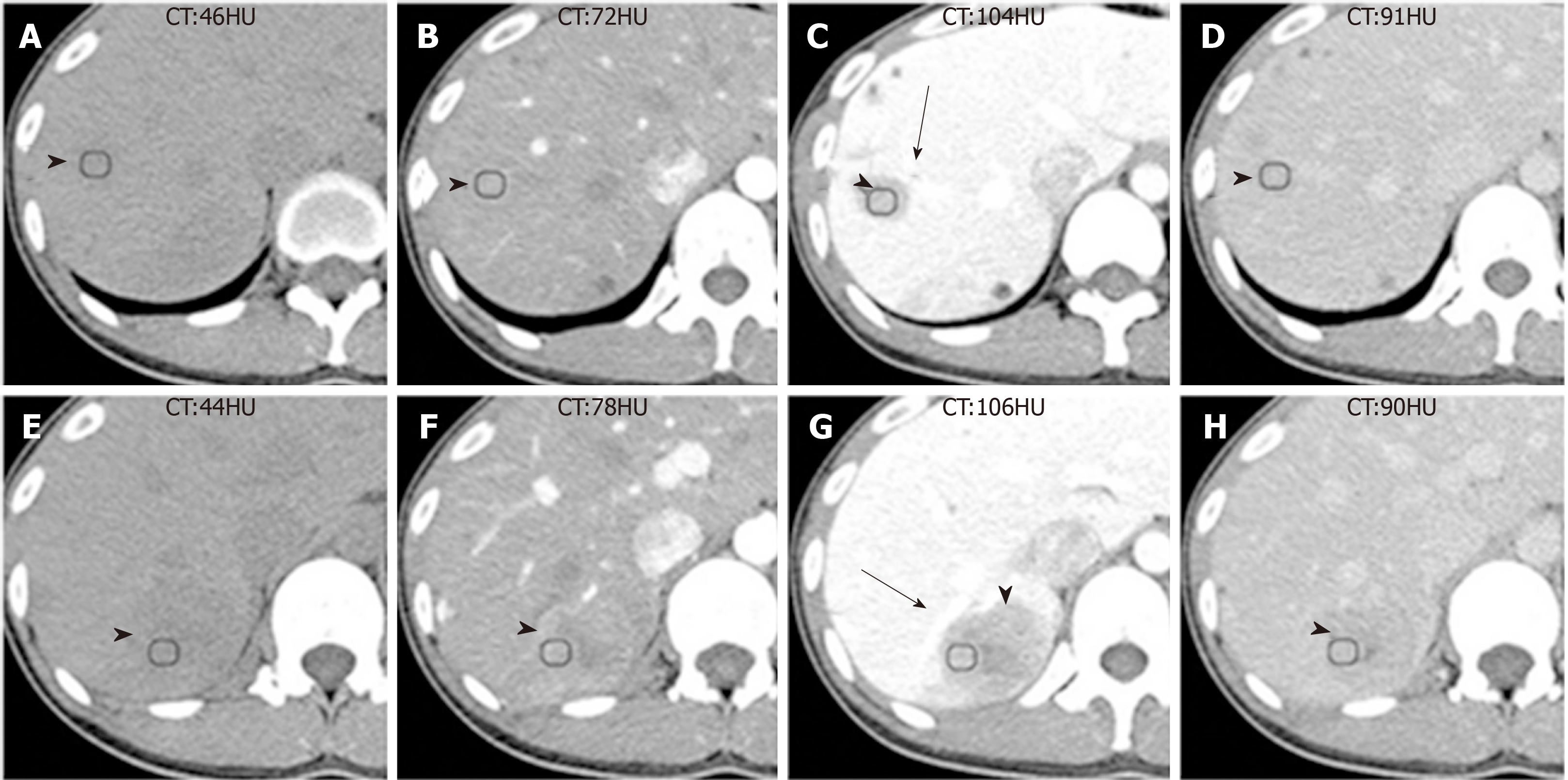
Figure 1 Computed tomography images of Case 1.
A and E: The unenhanced computed tomography (CT) images showed two well-defined hypodense masses; B-D, F-H: The triple-phase enhanced CT images showed both tumors had heterogeneous sustained hypoenhancement with internal necrosis. As the enhanced scan time was extended, the range of low density components within the mass decreased. Both tumors were close to the portal vein branch (black arrows) and always exhibited hypoattenuation relative to the surrounding liver parenchyma. Inflammatory pseudotumor-like follicular dendritic cell tumors (black arrowheads) from the upper segment of the right posterior lobe of the liver were present in a 31-year-old Chinese woman. CT: Computed tomography.
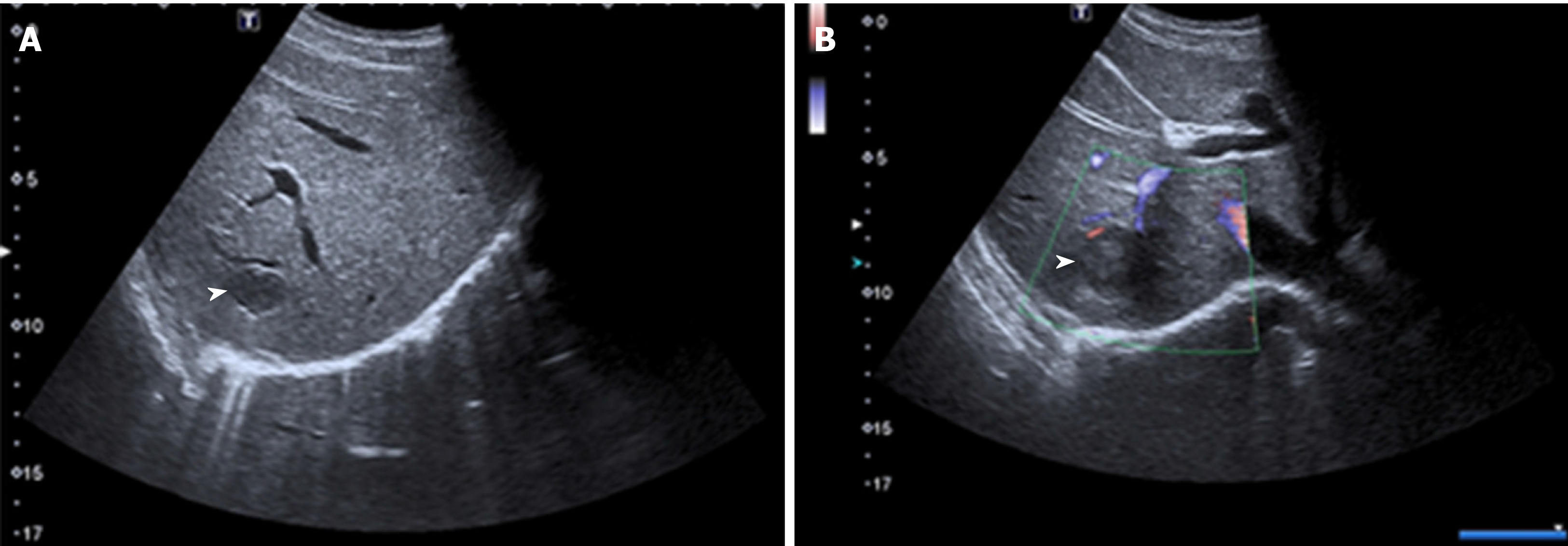
Figure 2 Unenhanced ultrasound images of Case 1.
A and B: Unenhanced ultrasound revealed two heterogeneous hypoechoic lesions without visualized internal blood flow. Inflammatory pseudotumor-like follicular dendritic cell tumors (black arrowheads) from the upper segment of the right posterior lobe were present in the liver of a 31-year-old Chinese woman.
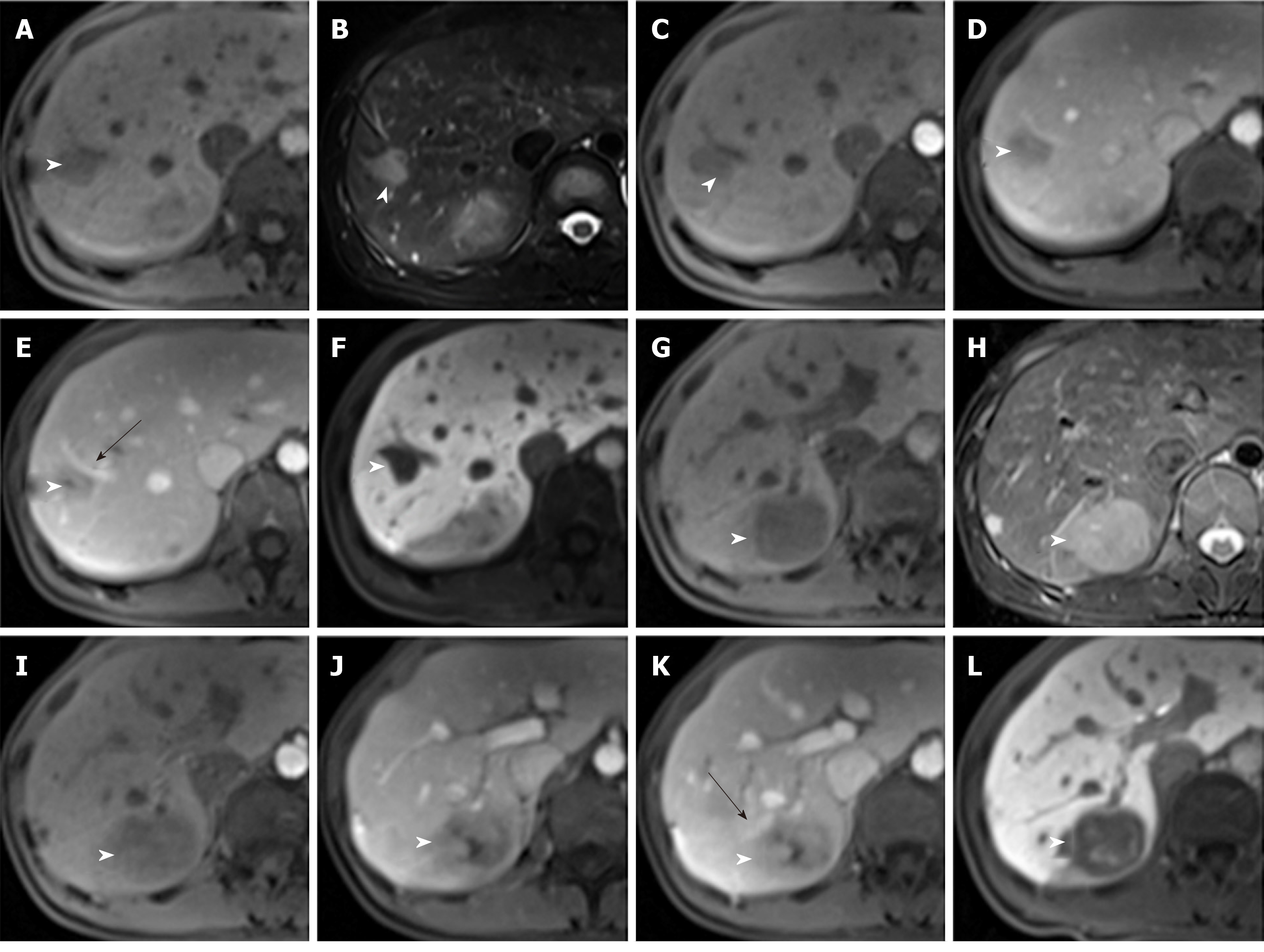
Figure 3 Magnetic resonance images of Case 1.
A and G: T1 weighted imaging (WI) showed two ovalular masses with heterogenous hypointense signals; B and H: T2WI showed two ovalular masses with heterogenous hyperintense signals; C-E, I-K: Triple-phase enhanced magnetic resonance images showed both tumors had heterogeneous sustained hypoenhancement with internal necrosis. As enhanced scan time was extended, the range of low density components within the mass decreased. Both tumors were close to the portal vein branch (black arrows) and always exhibited hypoattenuation relative to the surrounding liver parenchyma; F and L: The hepatobiliary phase exhibited both tumors with patchy hyperintense signal. Inflammatory pseudotumor-like follicular dendritic cell tumors (black arrowheads) from the upper segment of the right posterior lobe of the liver were present in a 31-year-old Chinese woman.
Figure 4 Unenhanced ultrasound images of Case 2.
A and B: Unenhanced ultrasound revealed one massive heterogeneous hypoecho lesion with internal blood flow located in the right lobe of the liver. Inflammatory pseudotumor-like follicular dendritic cell tumors (black arrowheads) from the right lobe of the liver were present in a 48-year-old Chinese man.
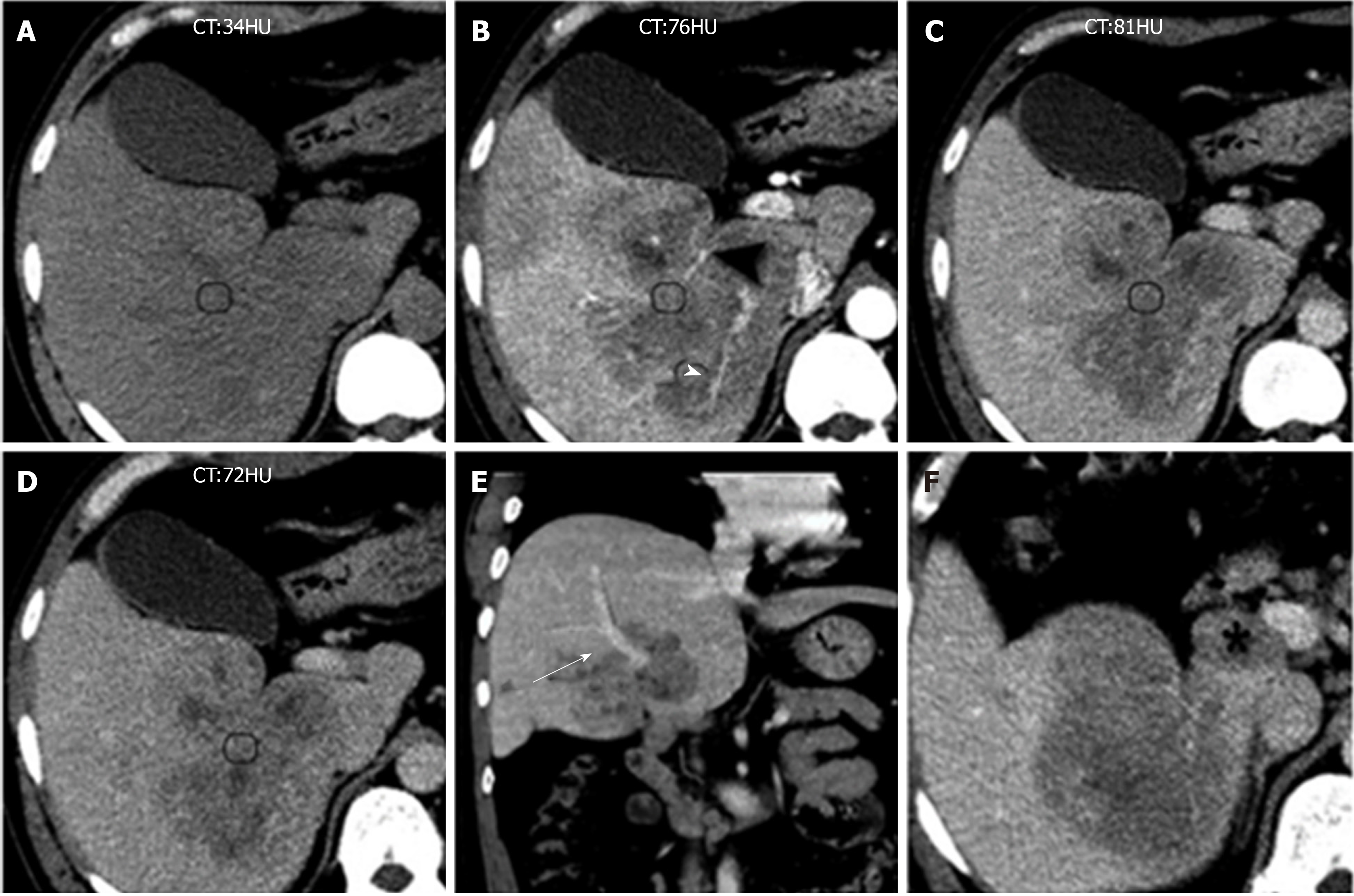
Figure 5 Computed tomography images of Case 2.
A: Unenhanced computed tomography (CT) image showing huge ill-defined heterogeneous hypodense masses; B-D: Multiphase enhanced CT images show a tumor with marked heterogeneous sustained hypoenhancement with internal necrosis. Engorged vascular structures (black arrowheads) were present in the mass; E: Coronal contrast-enhanced CT image showing the tumor near the portal vein (black arrow); and F: Axial contrast-enhanced CT image showing an enlarged lymph node seen in the hepatic hilar region (black star). Inflammatory pseudotumor-like follicular dendritic cell tumors from the right lobe of the liver were present in a 48-year-old Chinese man. CT: Computed tomography.
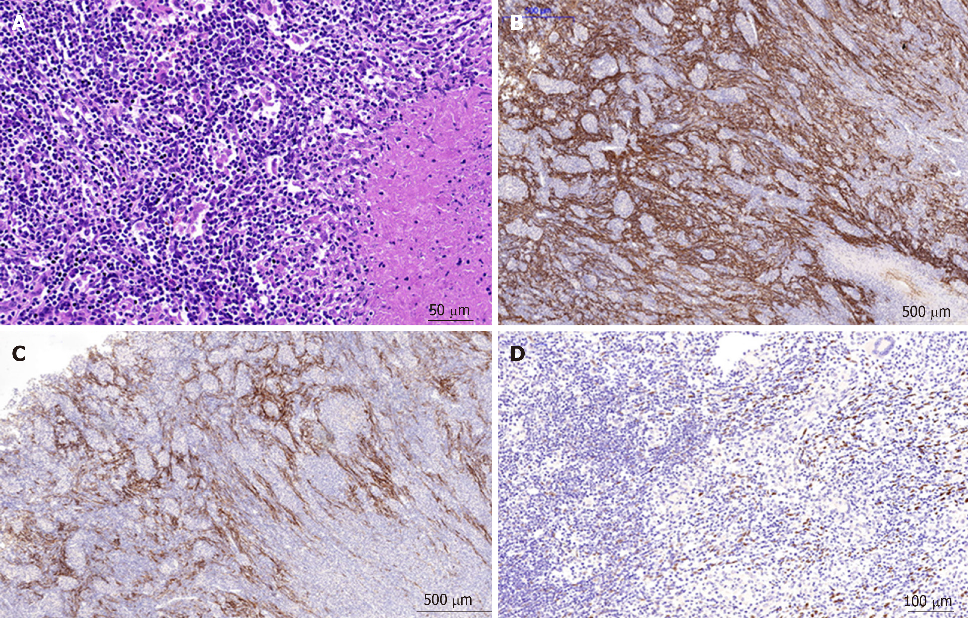
Figure 6 Inflammatory pseudotumor-like follicular dendritic cell tumor from the liver of a 31-year-old Chinese woman.
A: Histology by hematoxylin and eosin staining showed the tumor was composed of spindle cells, abundant small lymphocytes and plasmocytes, mixed with multifocal coagulative necrosis and granulomas; B: Immunohistochemical images showed positive staining for CD21; C: Immunohistochemical images showed positive staining for CD23; and D: Immunohistochemical images showed positive staining for Epstein-Barr-encoded-RNA (EBER). CD21, CD23, and EBER are specific biomarkers for diagnosing inflammatory pseudotumor-like follicular dendritic cell tumors.
- Citation: Li HL, Liu HP, Guo GWJ, Chen ZH, Zhou FQ, Liu P, Liu JB, Wan R, Mao ZQ. Imaging findings of inflammatory pseudotumor-like follicular dendritic cell tumors of the liver: Two case reports and literature review. World J Gastroenterol 2019; 25(45): 6693-6703
- URL: https://www.wjgnet.com/1007-9327/full/v25/i45/6693.htm
- DOI: https://dx.doi.org/10.3748/wjg.v25.i45.6693


