Copyright
©The Author(s) 2019.
World J Gastroenterol. Nov 28, 2019; 25(44): 6571-6578
Published online Nov 28, 2019. doi: 10.3748/wjg.v25.i44.6571
Published online Nov 28, 2019. doi: 10.3748/wjg.v25.i44.6571
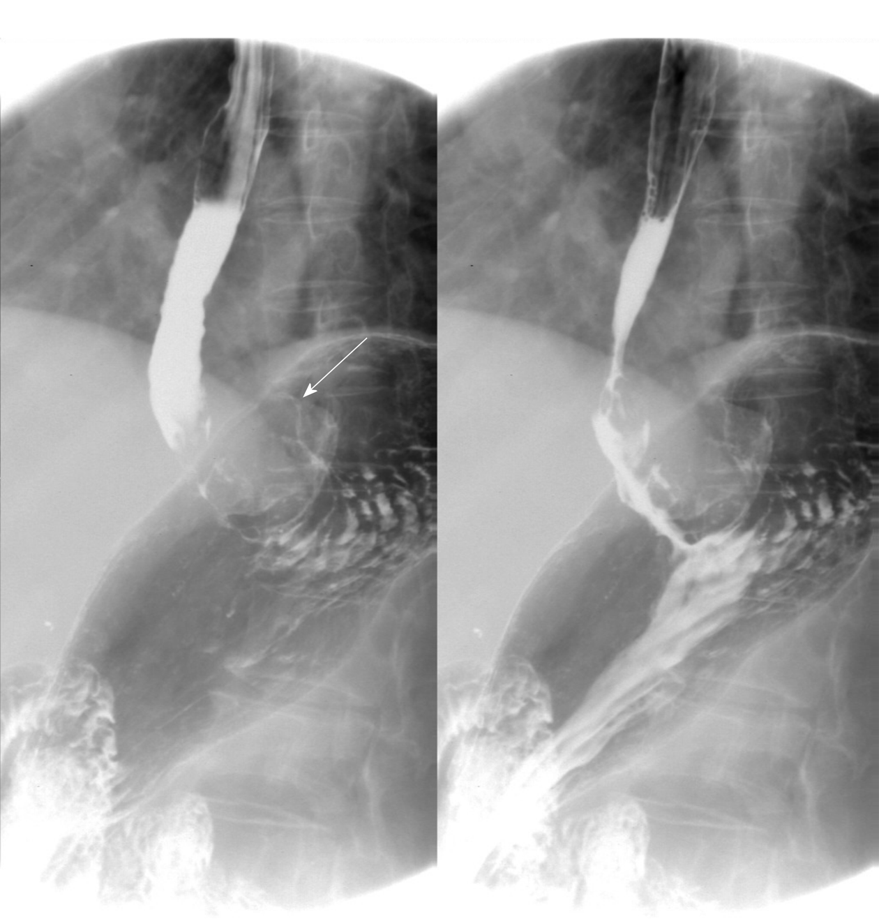
Figure 1 Digital gastrointestinal radiography showed that the tumor was located in the gastric cardia.
Thickening and rigidity of the gastric wall with no peristalsis were detected.
Figure 2 Computed tomography images.
A: Computed tomography plain scanning revealed an iso-or slight low-density tumor; B: The tumor showed heterogeneous enhancement in the arterial phase; C: Persistent enhancement was found in the portal vein phase.
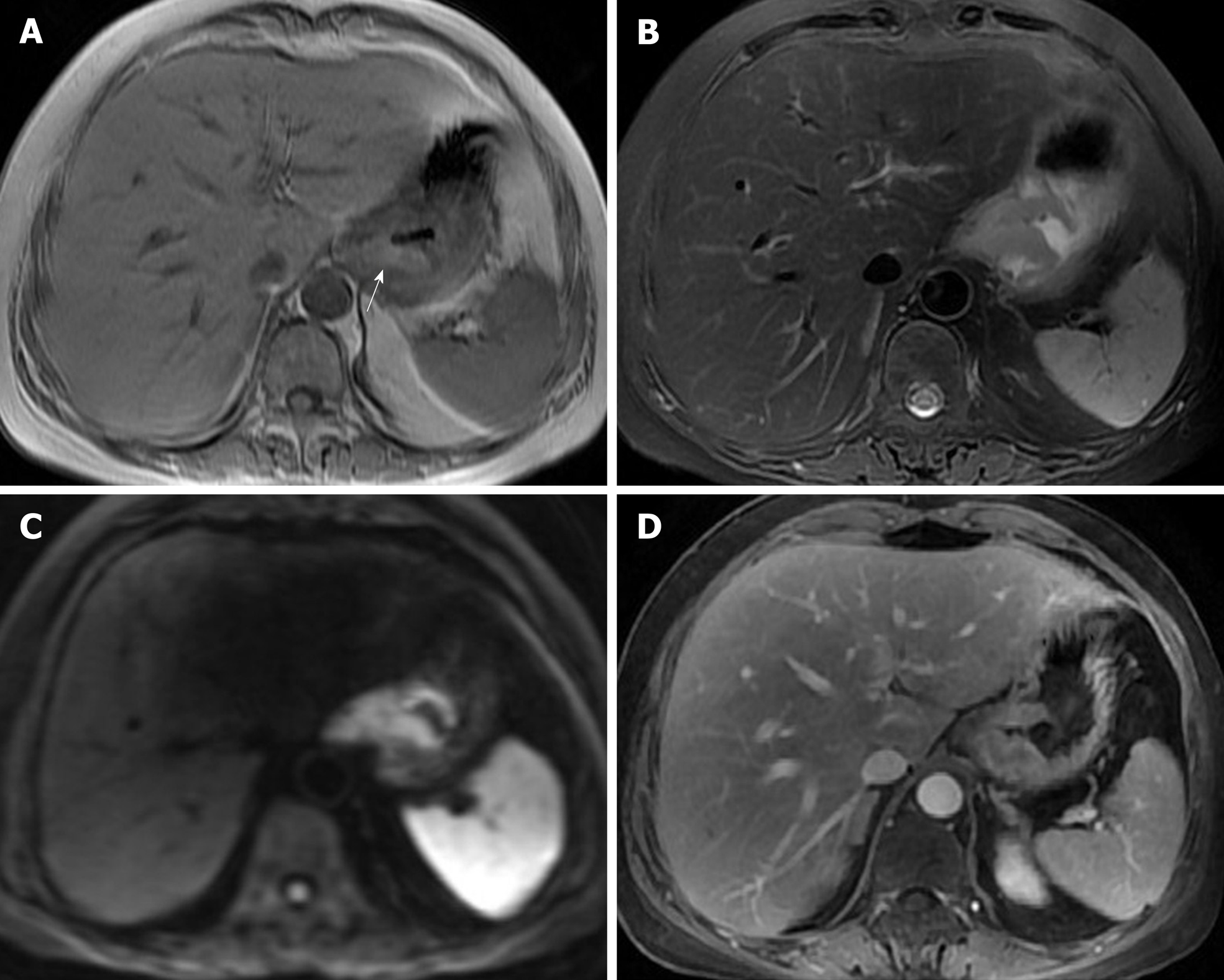
Figure 3 Magnetic resonance imaging images.
A: On magnetic resonance imaging (MRI), the mass exhibited a heterogeneous hyperintensity on T1-weighted imaging; B: On MRI, the mass exhibited a heterogeneous hypointensity on T2-weighted imaging; C: Diffusion weighted imaging showed hyperintense signal; D: On enhanced image, the lesion was obviously unevenly enhanced.
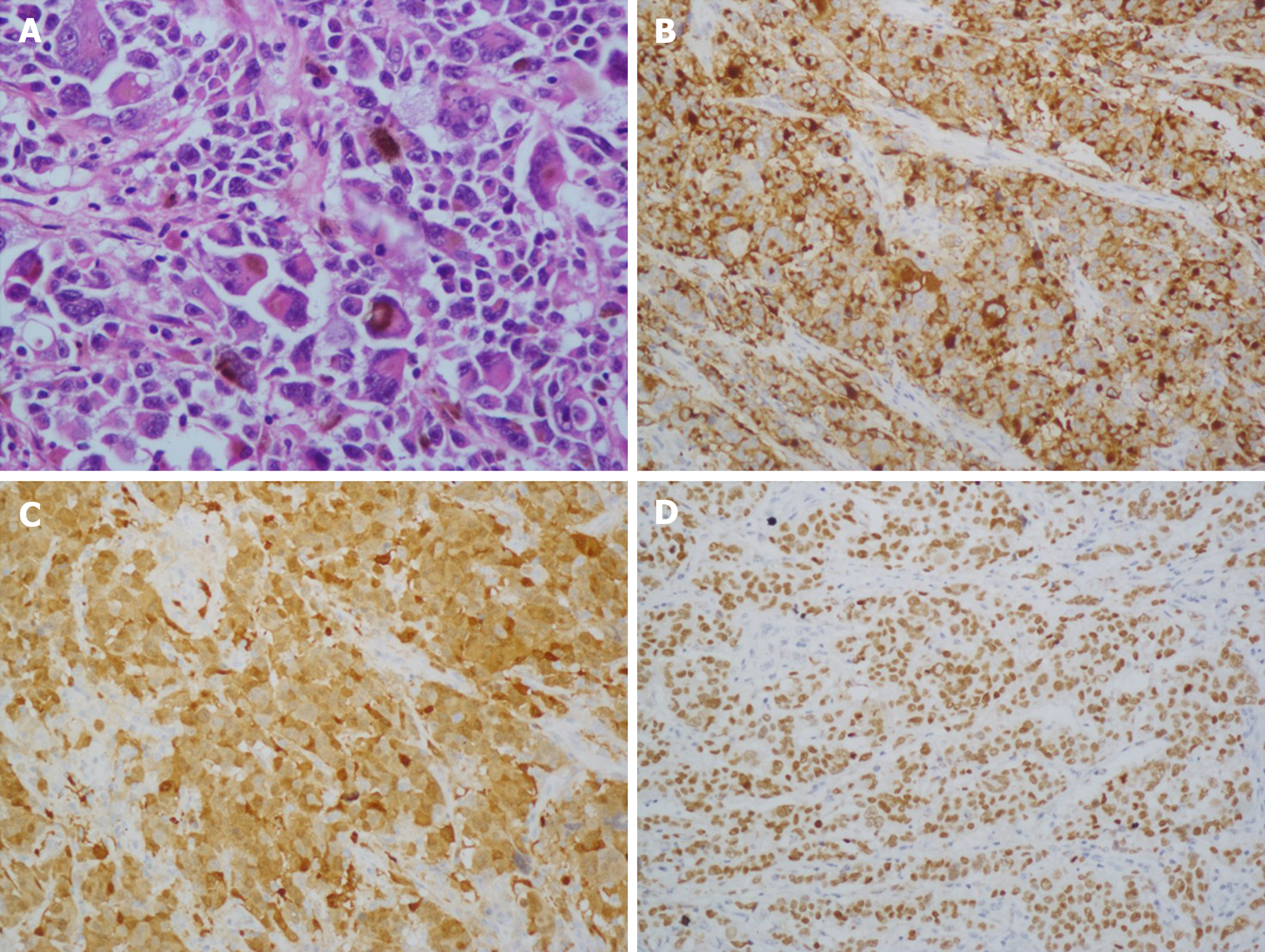
Figure 4 Histological and immunohistochemical images.
A: Hematoxylin and eosin staining (× 100) showed that the tumor cells were lymphocyte-like with pigmentation; B-D: Immunohistochemical staining showed that the primary gastric melanoma (PGM) was positive for HMB-45 (B; × 100), S100 (C; × 400), and MITF (D; × 100).
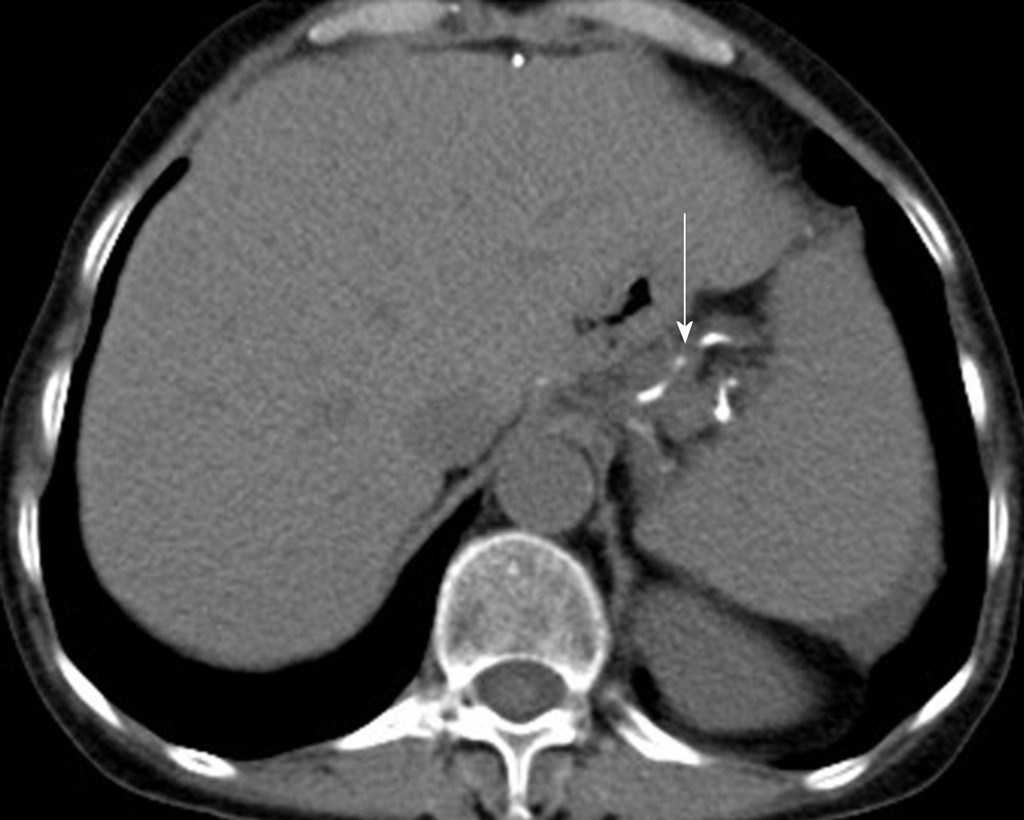
Figure 5 Re-examination 36 mo after total gastrectomy.
Computed tomography revealed that no recurrence or metastasis was observed. Surgical sutures were seen (arrow).
- Citation: Wang J, Yang F, Ao WQ, Liu C, Zhang WM, Xu FY. Primary gastric melanoma: A case report with imaging findings and 5-year follow-up. World J Gastroenterol 2019; 25(44): 6571-6578
- URL: https://www.wjgnet.com/1007-9327/full/v25/i44/6571.htm
- DOI: https://dx.doi.org/10.3748/wjg.v25.i44.6571


