Copyright
©The Author(s) 2019.
World J Gastroenterol. Nov 7, 2019; 25(41): 6190-6204
Published online Nov 7, 2019. doi: 10.3748/wjg.v25.i41.6190
Published online Nov 7, 2019. doi: 10.3748/wjg.v25.i41.6190
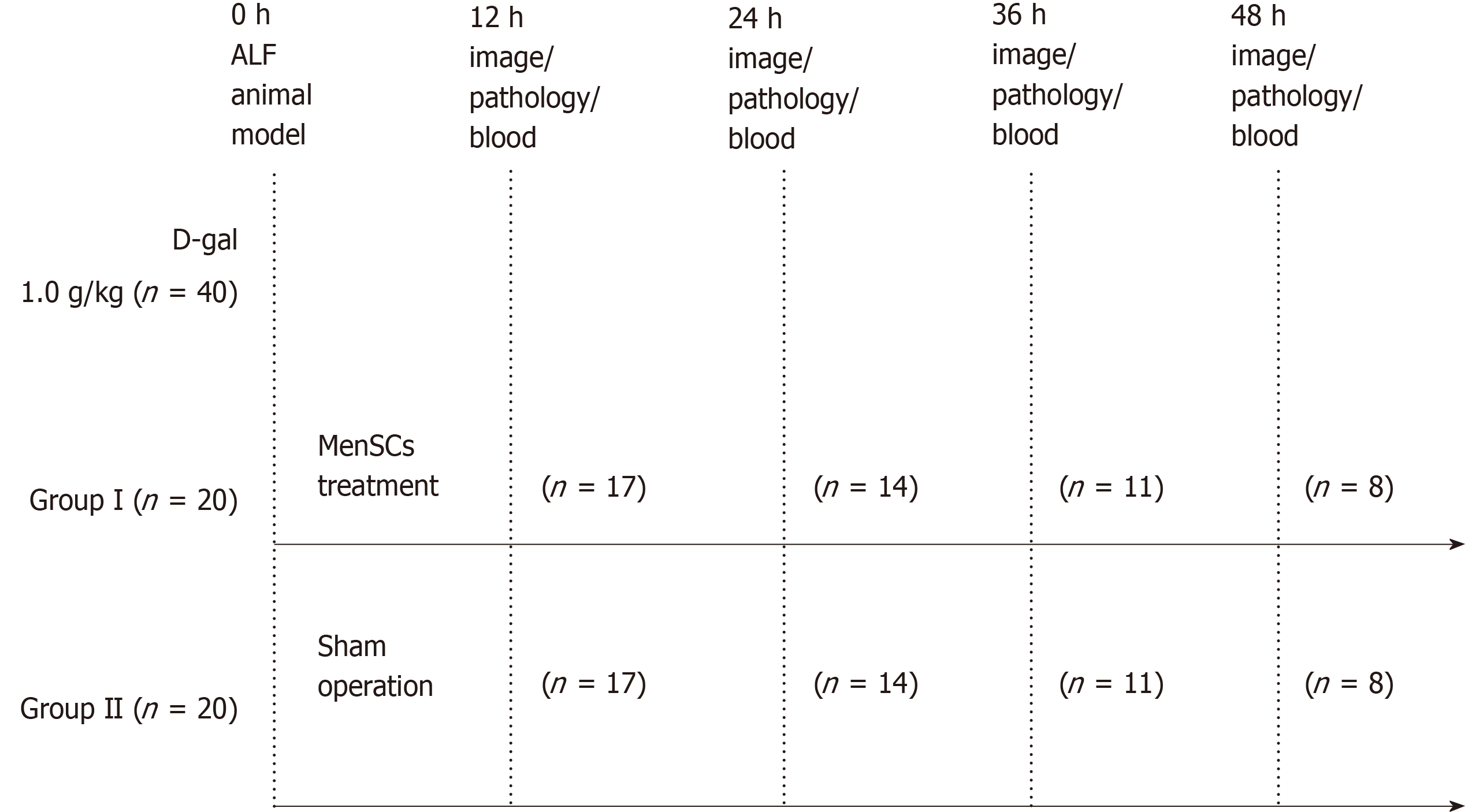
Figure 1 Experimental design.
Acute liver failure (ALF) was induced in forty animals with D-galactosamine (D-gal) at a dose of 1.0 g/kg. The treatment group (Group I, n = 20) received cell transplantation and the control group (Group II, n = 20) received a sham operation. Animals from both groups were sacrificed every 12 h. Blood samples were collected for biochemical analysis. Three major organs (the lungs, liver, and spleen) were isolated and immediately imaged with the In vivo Imaging System. Then liver tissues were collected for pathological examination. ALF: Acute liver failure; MenSCs: Menstrual blood stem cells; D-gai: D-galactosamine; IVIS: In vivo Imaging System.
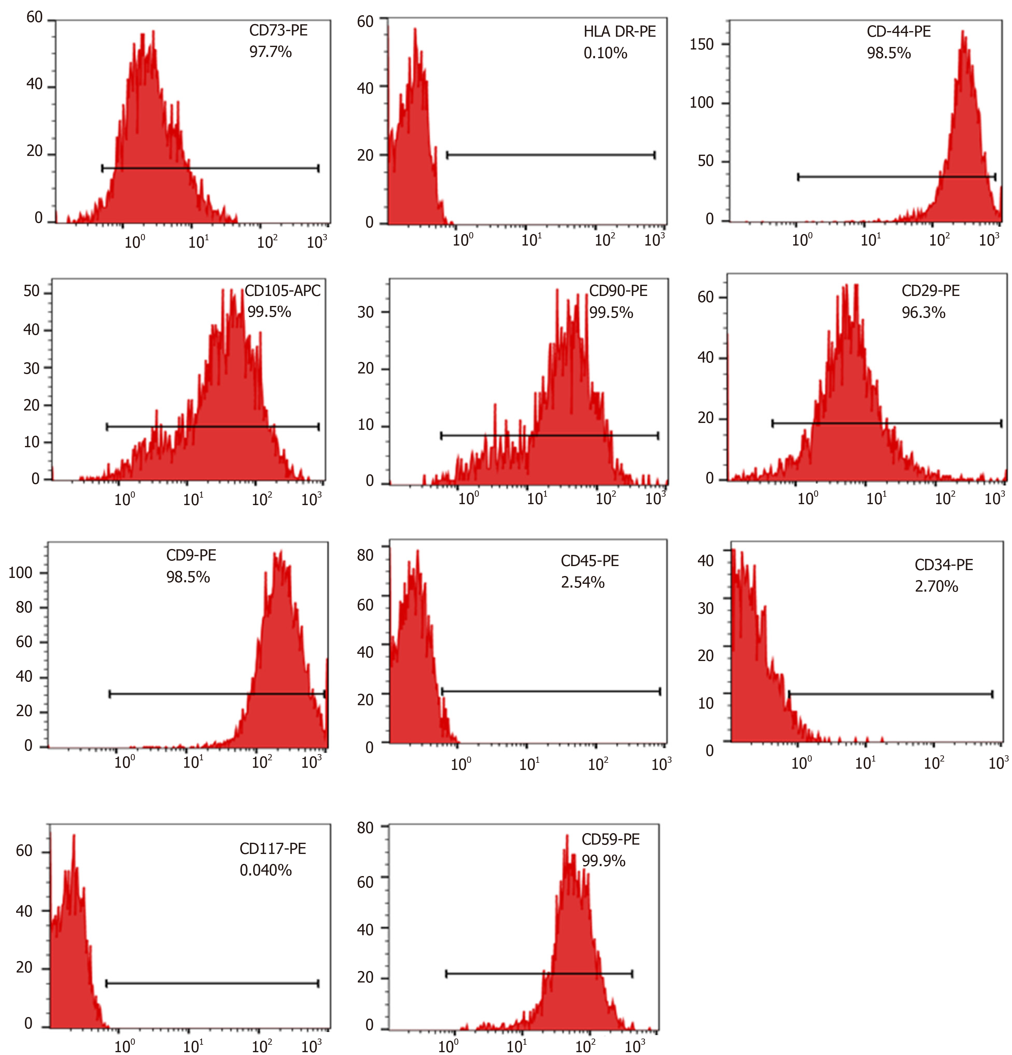
Figure 2 Phenotypic profile of menstrual blood stem cells isolated from menstrual blood.
Surface markers of cultured menstrual blood stem cells (MenSCs) were analyzed using a flowcytometer at the third and fifth passages. MenSCs: Menstrual blood stem cells.
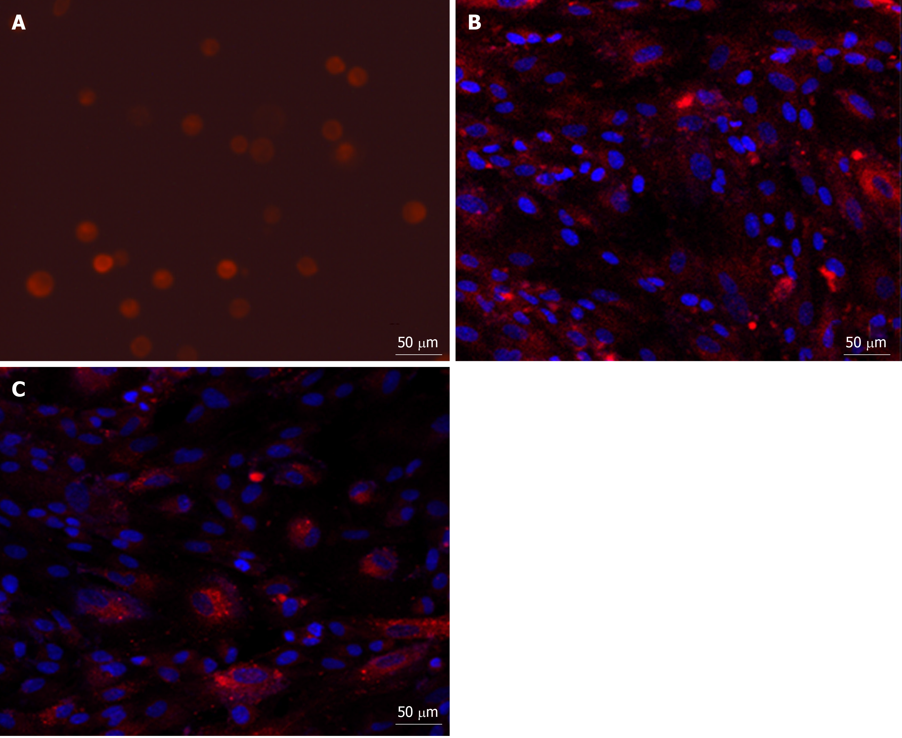
Figure 3 Fluorescence micrograph of PKH26-labelled menstrual blood stem cells.
A: The third passage of PKH26-labelled menstrual blood stem cells (MenSCs) was resuspended and exhibited red fluorescence (×20); B: When the labelled MenSCs were cultured in vitro, most of the cells were able to grow adhering to the wall (×20); C: After serial passages, the fluorescent signal intensity from the cells decreased with time (×20, passage 8). MenSCs: Menstrual blood stem cells.
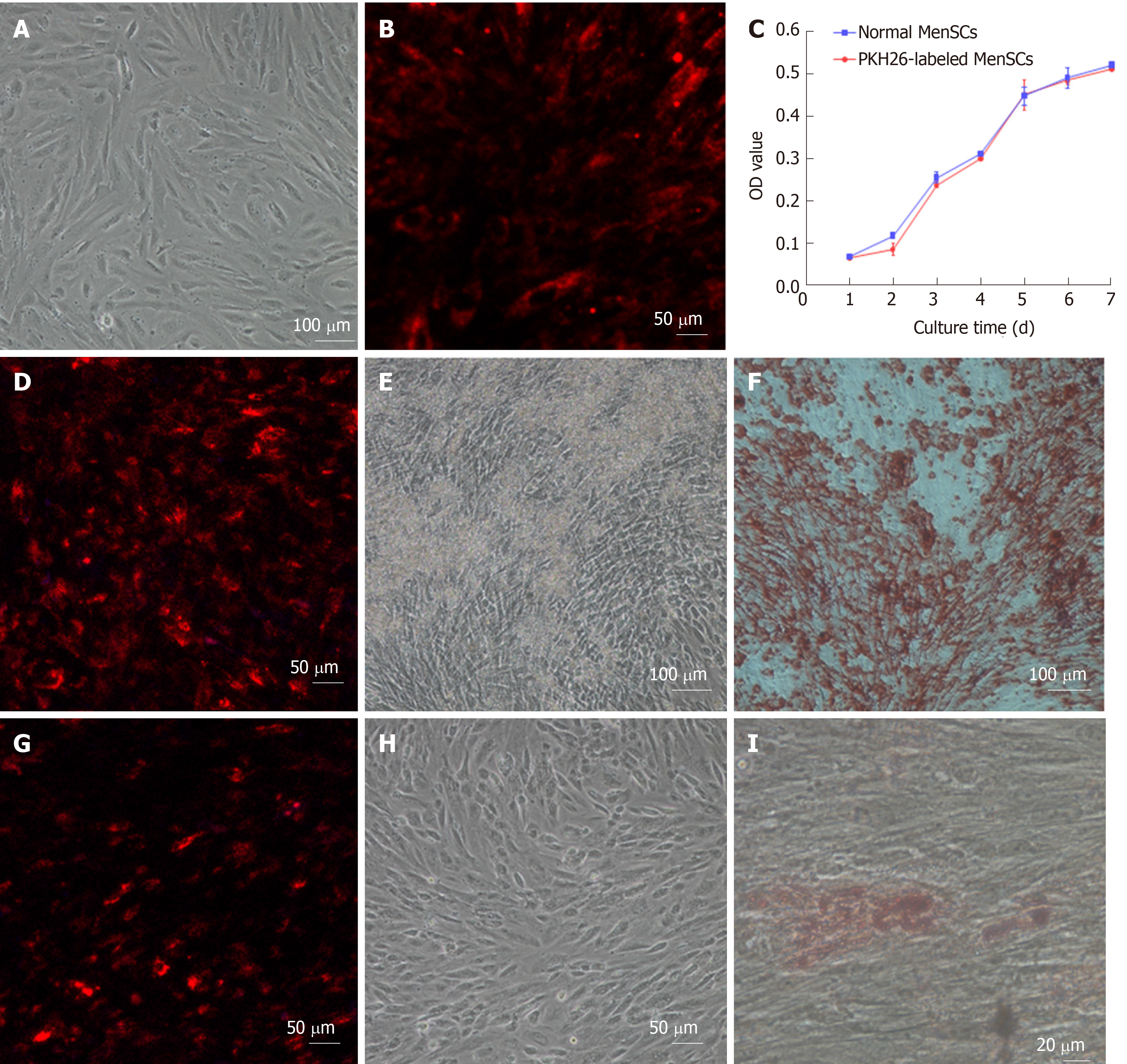
Figure 4 Effect of PKH26 on the cytomorphology, viability and multipotential differentiation of menstrual blood stem cells.
A: Morphology of the fifth passage of menstrual blood stem cells (MenSCs) without PKH26 labelling (× 10); B: the fifth passage of MenSCs with PKH26 labelling after 3 d (× 20); C: Viability of PKH26-labelled MenSCs and non-labelled normal MenSCs; D: Osteogenic differentiation of PKH26-labelled MenSCs under a confocal microscope (× 20); E: Bone nodules formed by differentiated osteocytes were visualized (× 10); F: PKH26-labelled MenSCs underwent osteogenic differentiation after alizarin red S staining (× 10); G: Adipogenic differentiation of PKH26-labelled MenSCs under a confocal microscope (20 ×); H: Differentiated adipocytes were visualized before oil red O staining (× 10); I: Differentiated adipocytes contained lipid droplets after oil red O staining (× 40). MenSCs: Menstrual blood stem cells.
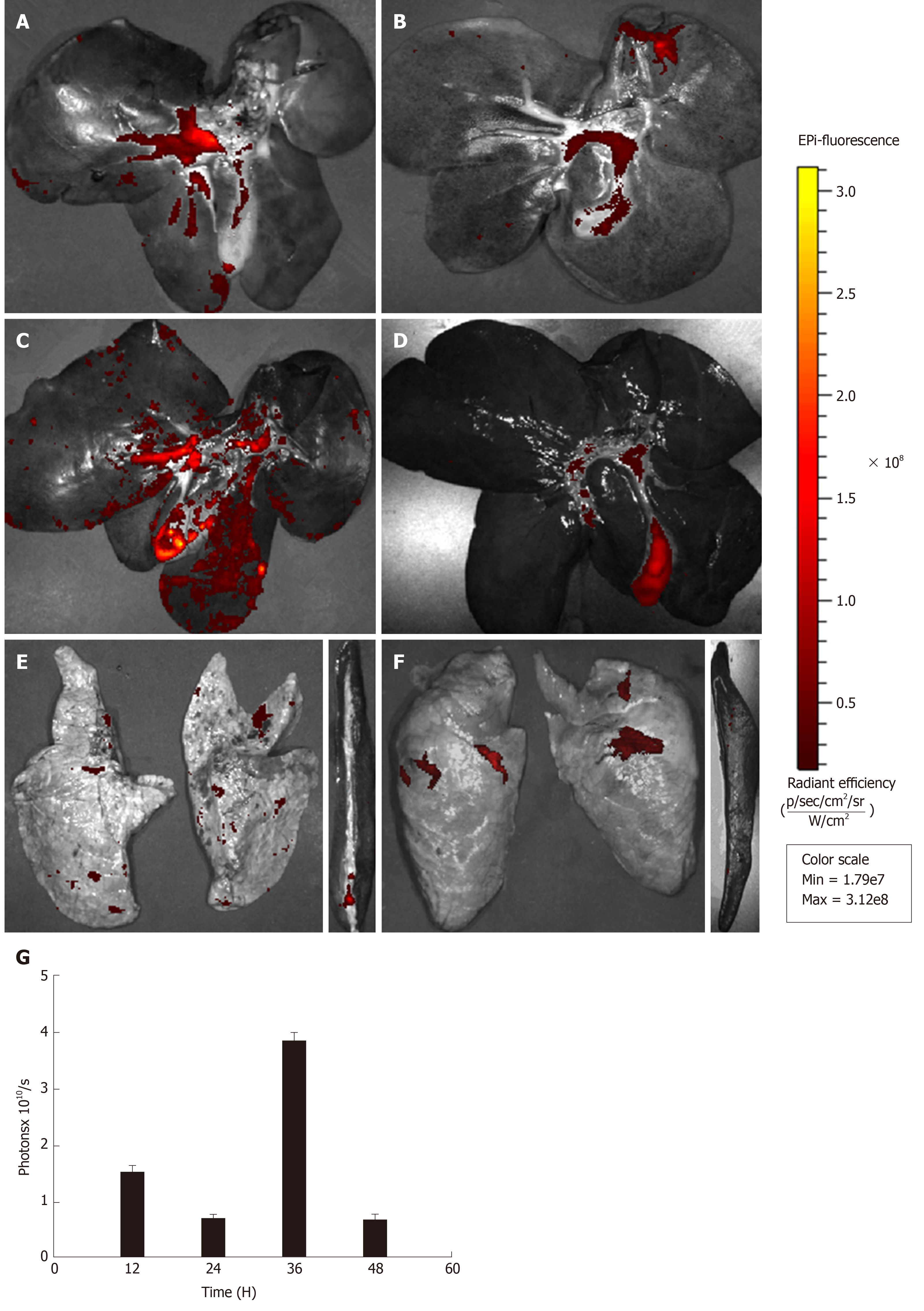
Figure 5 Ex vivo images showing the dynamic biodistribution of transplanted PKH26-labelled menstrual blood stem cells in different organs by the in vivo imaging system.
A: At 12 h after transplantation, strong red fluorescence emitted from PKH26-labelled menstrual blood stem cells (MenSCs) was detected in the liver; B: The fluorescence intensity of the liver weakened at 24 h; C: The fluorescence intensity of the liver increased notably at 36 h; D: Finally, the signal faded after 48 h; E: At 24 h after transplantation, a weak signal was detected in the lungs and the spleen; F: At 48 h after transplantation, a weak signal was also detected in the lungs, but it was seldom observed in the spleen; G: A graph showing the total photon emission efficiency in the liver at all time points. MenSCs: Menstrual blood stem cells; IVIS: In vivo Imaging System.
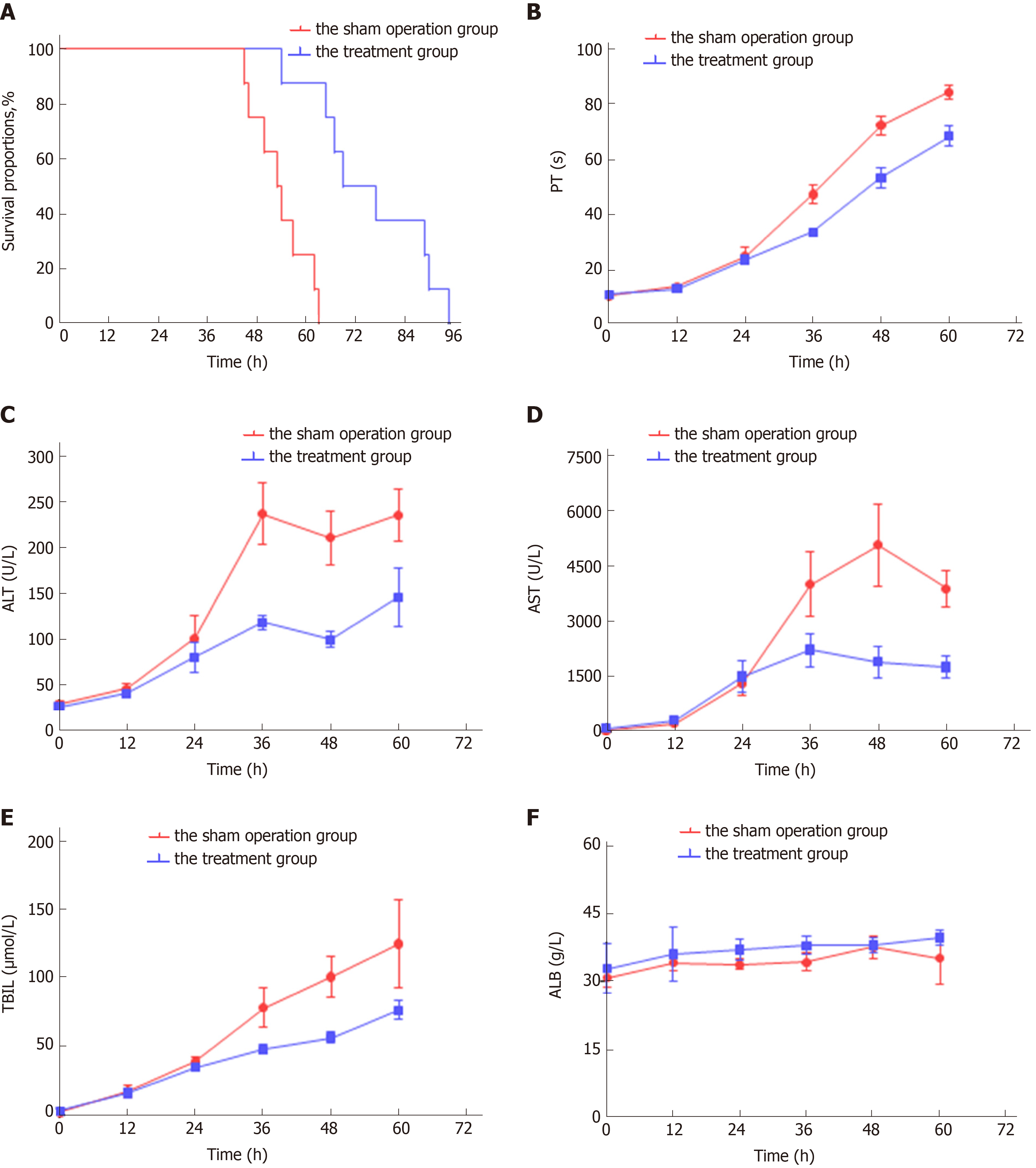
Figure 6 Efficacy of intraportally transplanted menstrual blood stem cells in treating pigs with acute liver failure.
A: Survival time of the treatment group and the sham operation group (P < 0.001); B: Dynamic changes in the biochemical in the two groups; C: Dynamic changes in the hematological parameters in the two groups. MenSCs: Menstrual blood stem cells; ALF: Acute liver failure; PT: Prothrombin time; ALT: Alanine aminotransferase; AST: Aspartate aminotransferase; TBIL: Total bilirubin; ALB: Albumin.
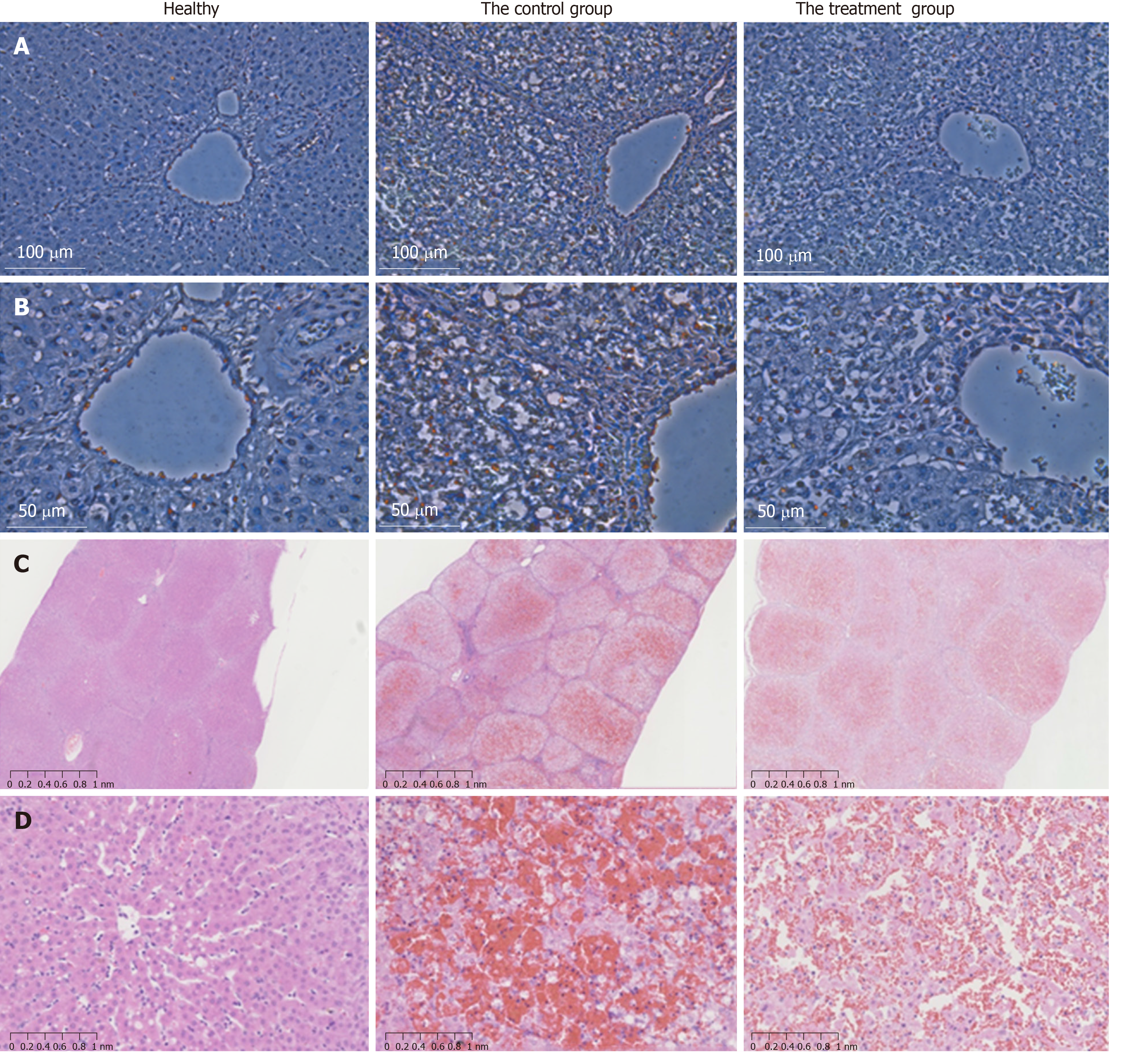
Figure 7 TUNEL assay and H&E staining of liver tissues after acute liver failure induction.
A: Apoptotic hepatocytes were stained red. At 36 h after ALF induction, the number of apoptotic cells was significantly lower in the menstrual blood stem cell (MenSC) treatment group than in the control group (× 20); B: High magnification images were shown (× 40); C: The livers of the ALF pigs without MenSC treatment showed a typical ALF histology with extensive haemorrhage, hepatocyte necrosis and adipose degeneration at 48 h. In contrast, ALF pigs with MenSC transplantation showed increased numbers of remaining hepatocytes and hepatic parenchymal cells (× 2); D: High magnification images were shown (× 20). MenSC treatment alleviated the progression of liver injury. ALF: acute liver failure; MenSC: menstrual blood stem cell.
- Citation: Cen PP, Fan LX, Wang J, Chen JJ, Li LJ. Therapeutic potential of menstrual blood stem cells in treating acute liver failure. World J Gastroenterol 2019; 25(41): 6190-6204
- URL: https://www.wjgnet.com/1007-9327/full/v25/i41/6190.htm
- DOI: https://dx.doi.org/10.3748/wjg.v25.i41.6190


