Copyright
©The Author(s) 2019.
World J Gastroenterol. Jan 28, 2019; 25(4): 485-497
Published online Jan 28, 2019. doi: 10.3748/wjg.v25.i4.485
Published online Jan 28, 2019. doi: 10.3748/wjg.v25.i4.485
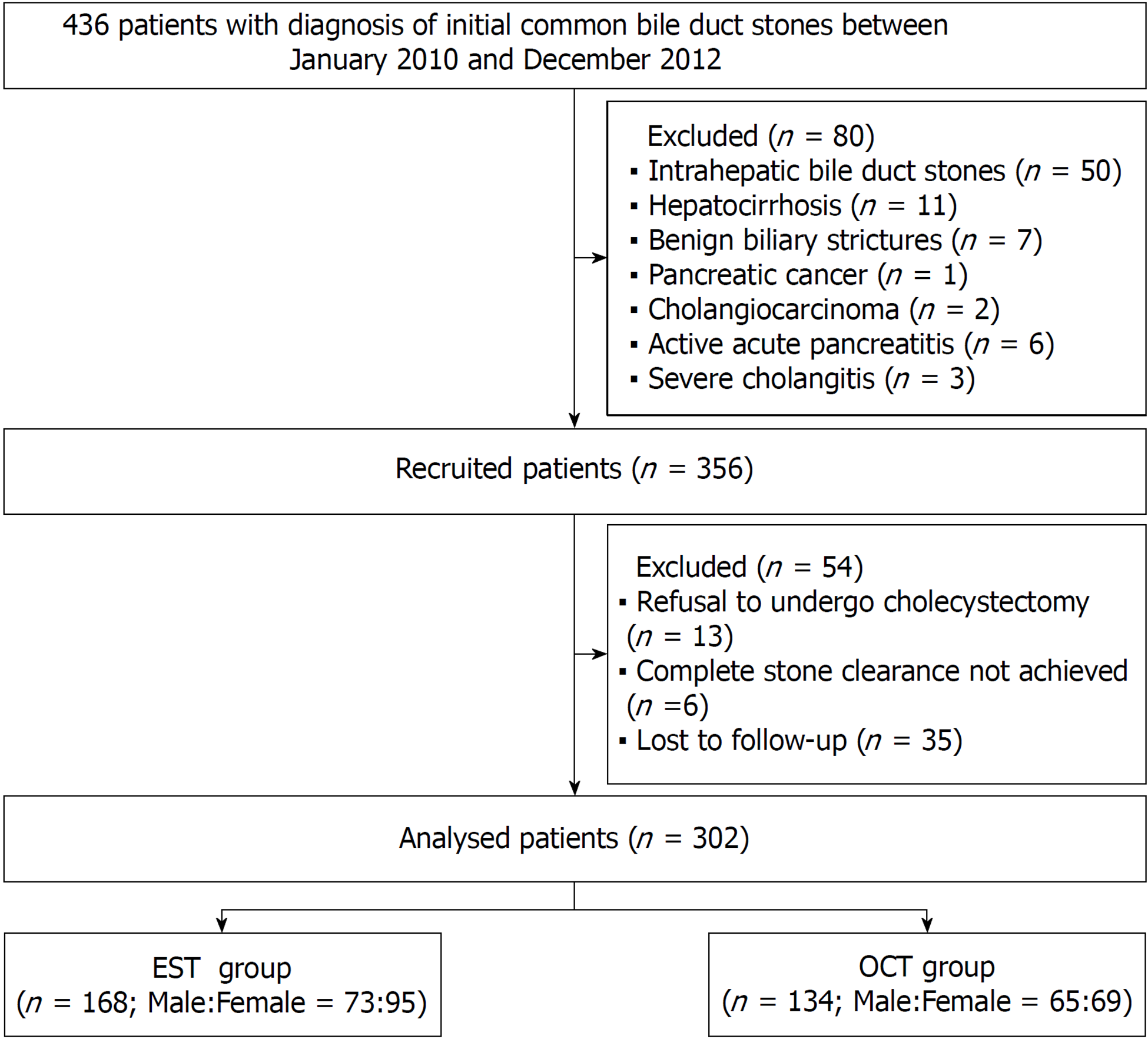
Figure 1 Flow diagram of patient selection.
EST: Endoscopic sphincterotomy; OCT: Open choledochotomy.
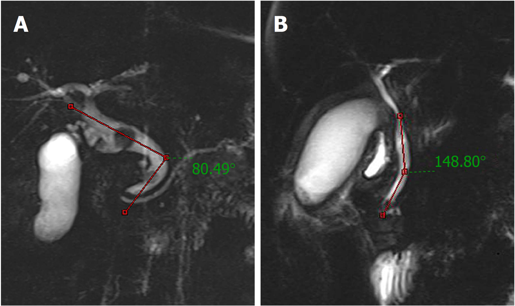
Figure 2 Distal common bile duct angulation measured based on magnetic resonance cholangiopancreatography.
A: A case with acute distal common bile duct angulation (< 145°); B: A case without acute distal common bile duct angulation (> 145°).
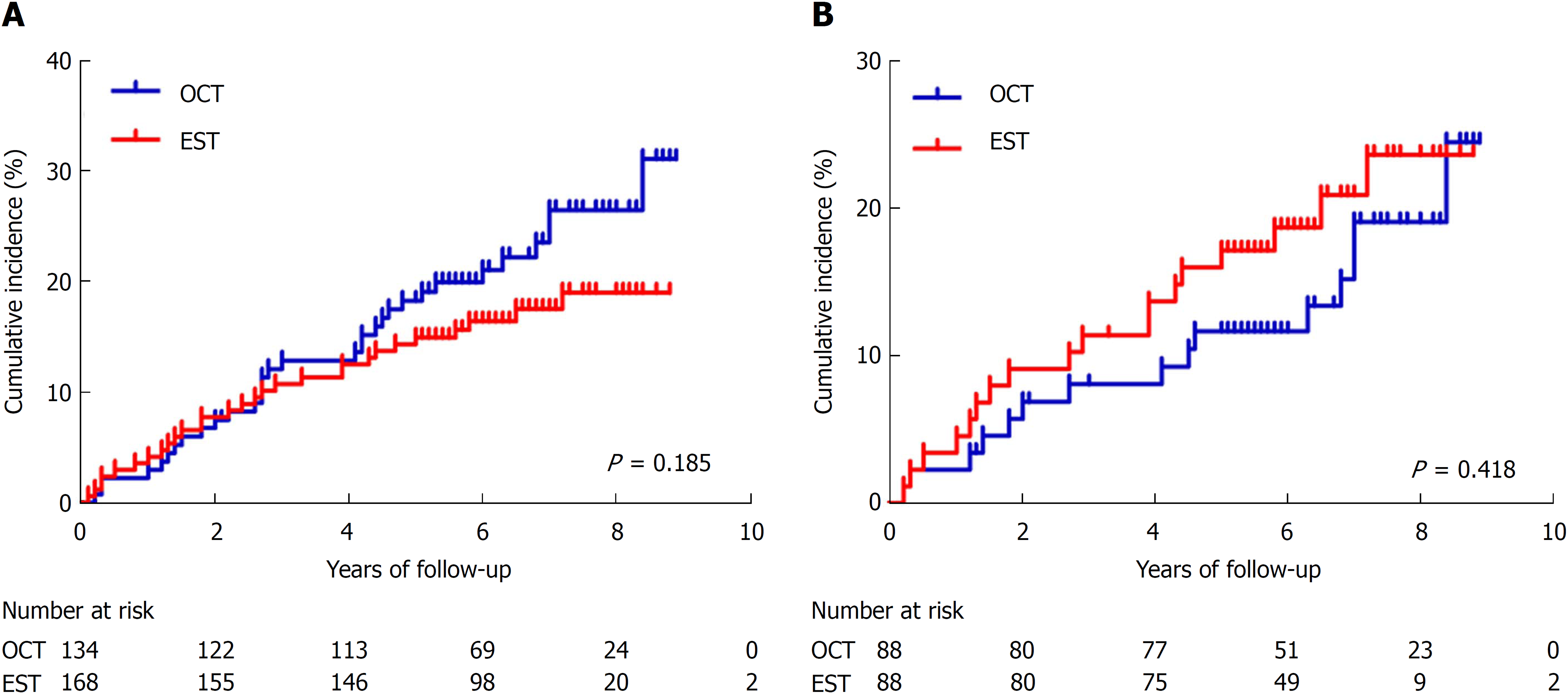
Figure 3 Kaplan-Meier analysis with the log-rank test for recurrence of common bile duct stones in the endoscopic sphincterotomy and open choledochotomy groups.
A: Before matching; B: After matching. EST: Endoscopic sphincterotomy; OCT: Open choledochotomy.
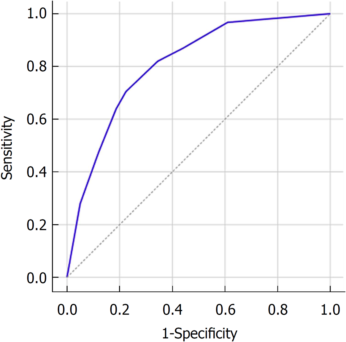
Figure 4 Receiver operating characteristic curve for the logistic regression model predicting recurrence of common bile duct stones.
This included common bile duct diameter ≥ 15 mm, multiple common bile duct stones, and distal common bile duct angle ≤ 145°. AUC = 0.81, 95%CI: 0.76-0.87, P < 0.001.
- Citation: Zhou XD, Chen QF, Zhang YY, Yu MJ, Zhong C, Liu ZJ, Li GH, Zhou XJ, Hong JB, Chen YX. Outcomes of endoscopic sphincterotomy vs open choledochotomy for common bile duct stones. World J Gastroenterol 2019; 25(4): 485-497
- URL: https://www.wjgnet.com/1007-9327/full/v25/i4/485.htm
- DOI: https://dx.doi.org/10.3748/wjg.v25.i4.485


