Copyright
©The Author(s) 2019.
World J Gastroenterol. Sep 14, 2019; 25(34): 5082-5096
Published online Sep 14, 2019. doi: 10.3748/wjg.v25.i34.5082
Published online Sep 14, 2019. doi: 10.3748/wjg.v25.i34.5082
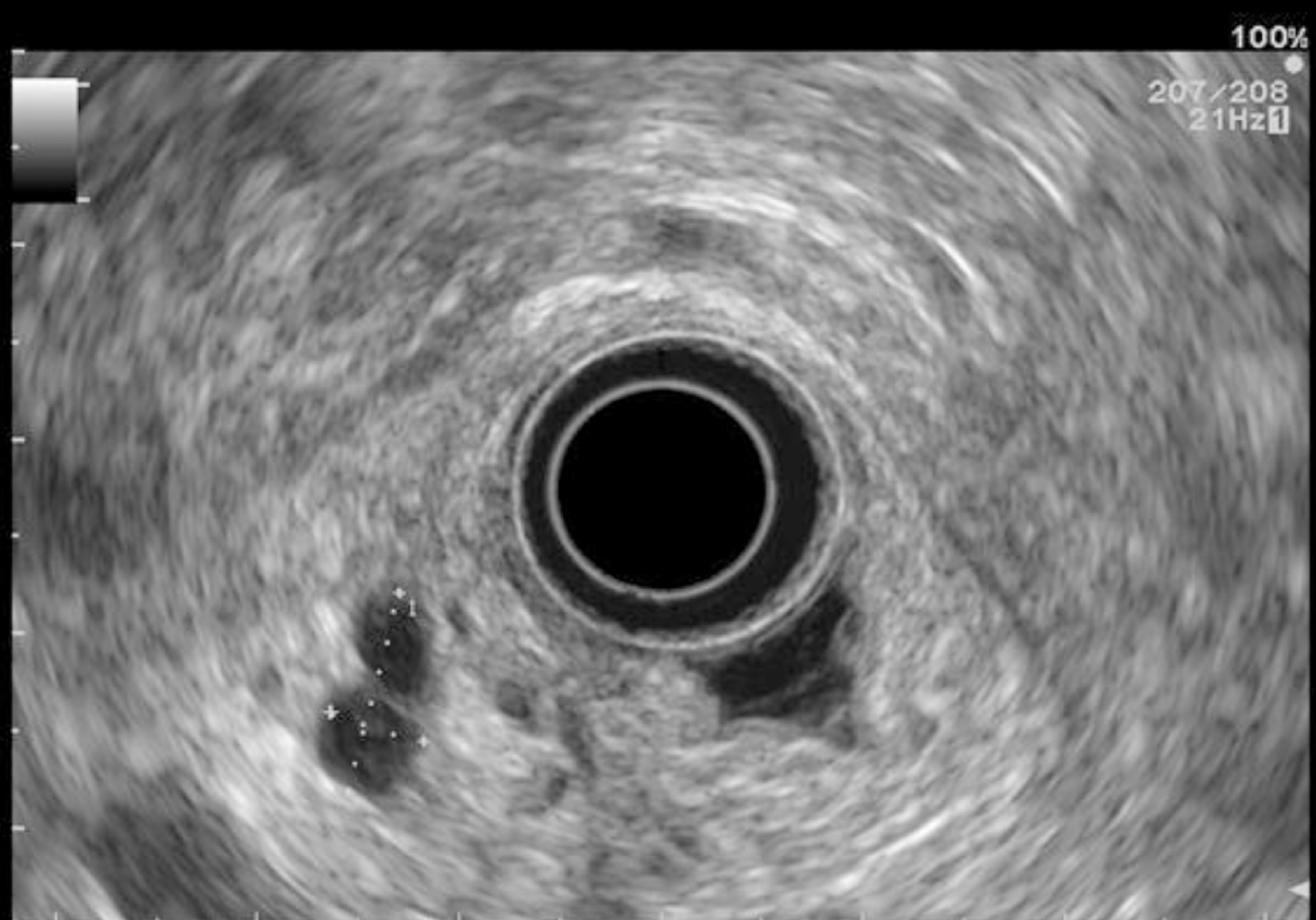
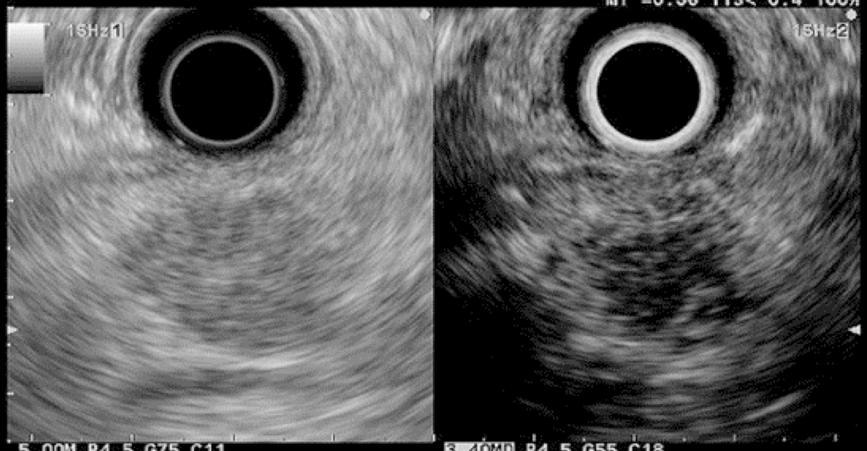
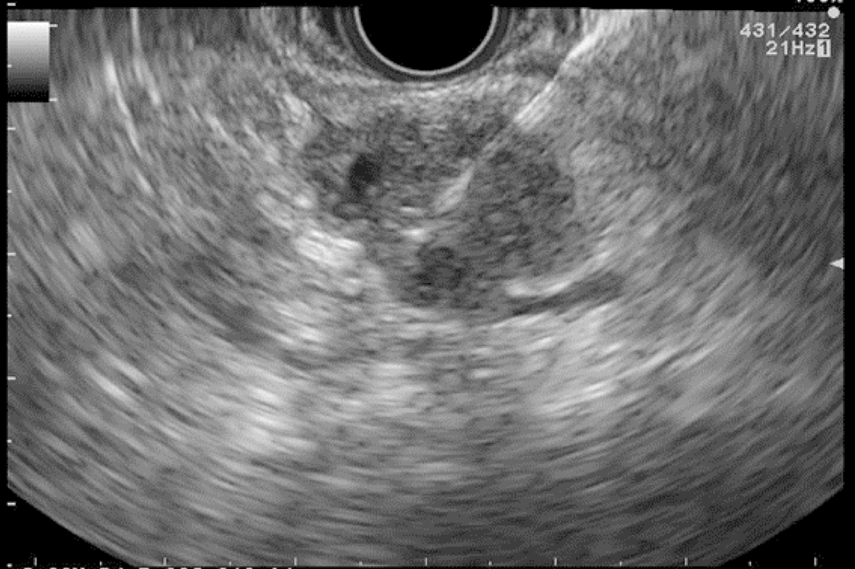
Figure 1 Pancreatic cystic lesion: Intraductal papillary mucinous neoplasm.
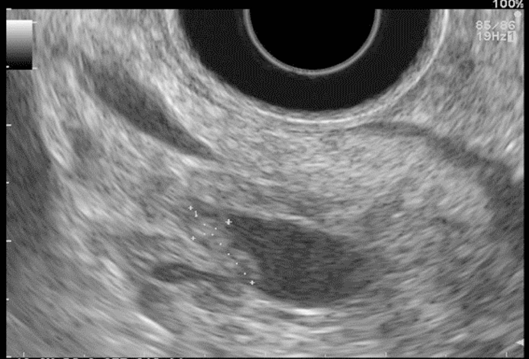
Figure 2 Intraductal papillary mucinous neoplasm with mural nodule.
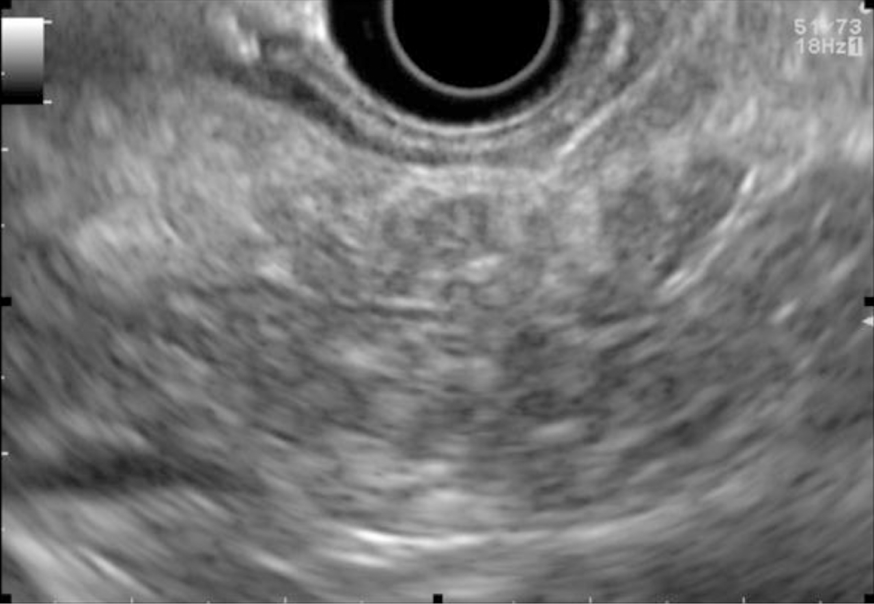
Figure 3 Chronic-pancreatitis-like parenchymal changes.
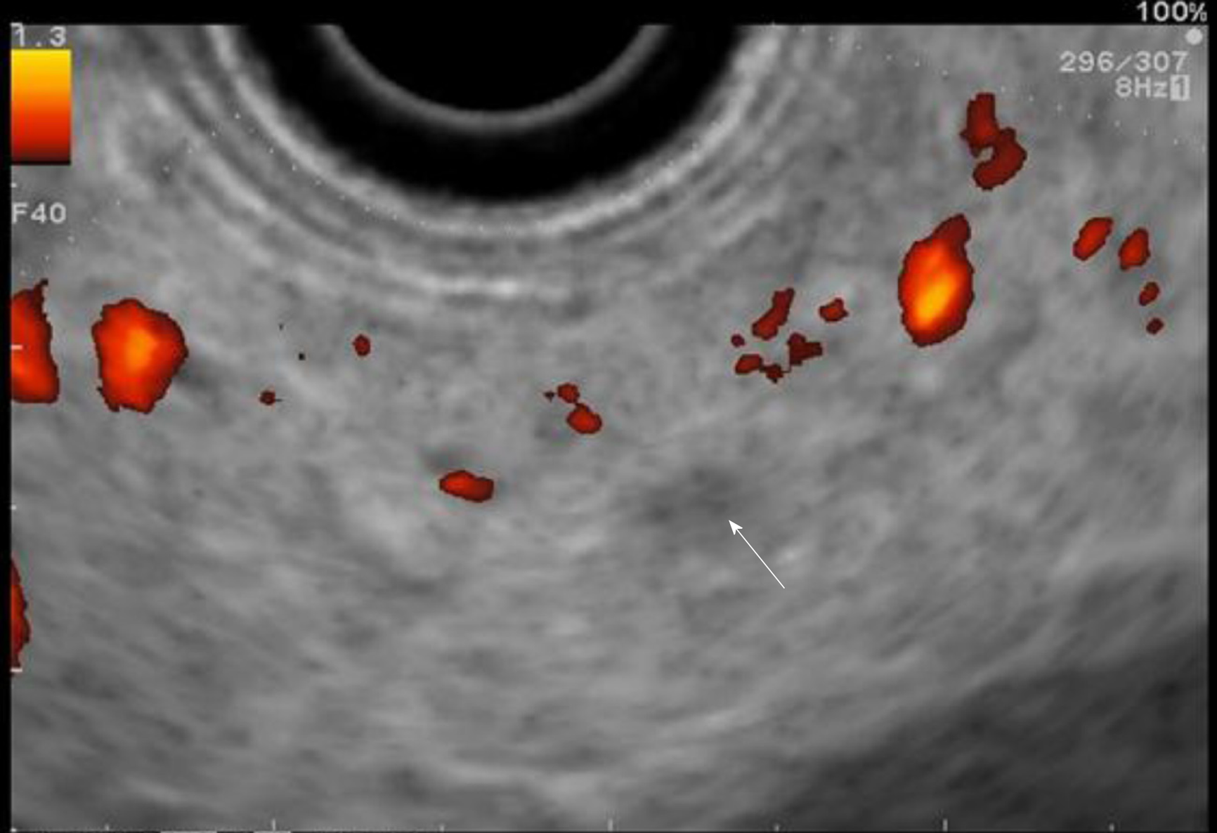
Figure 4 Small hypoechoic nodule.
Figure 5 Contrast-enhanced harmonic in endoscopic ultrasound: Hypoechoic suspect nodule.
Figure 6 Fine needle aspiration of a solid pancreatic lesion.
- Citation: Lorenzo D, Rebours V, Maire F, Palazzo M, Gonzalez JM, Vullierme MP, Aubert A, Hammel P, Lévy P, Mestier L. Role of endoscopic ultrasound in the screening and follow-up of high-risk individuals for familial pancreatic cancer. World J Gastroenterol 2019; 25(34): 5082-5096
- URL: https://www.wjgnet.com/1007-9327/full/v25/i34/5082.htm
- DOI: https://dx.doi.org/10.3748/wjg.v25.i34.5082


