Copyright
©The Author(s) 2019.
World J Gastroenterol. Aug 28, 2019; 25(32): 4764-4778
Published online Aug 28, 2019. doi: 10.3748/wjg.v25.i32.4764
Published online Aug 28, 2019. doi: 10.3748/wjg.v25.i32.4764
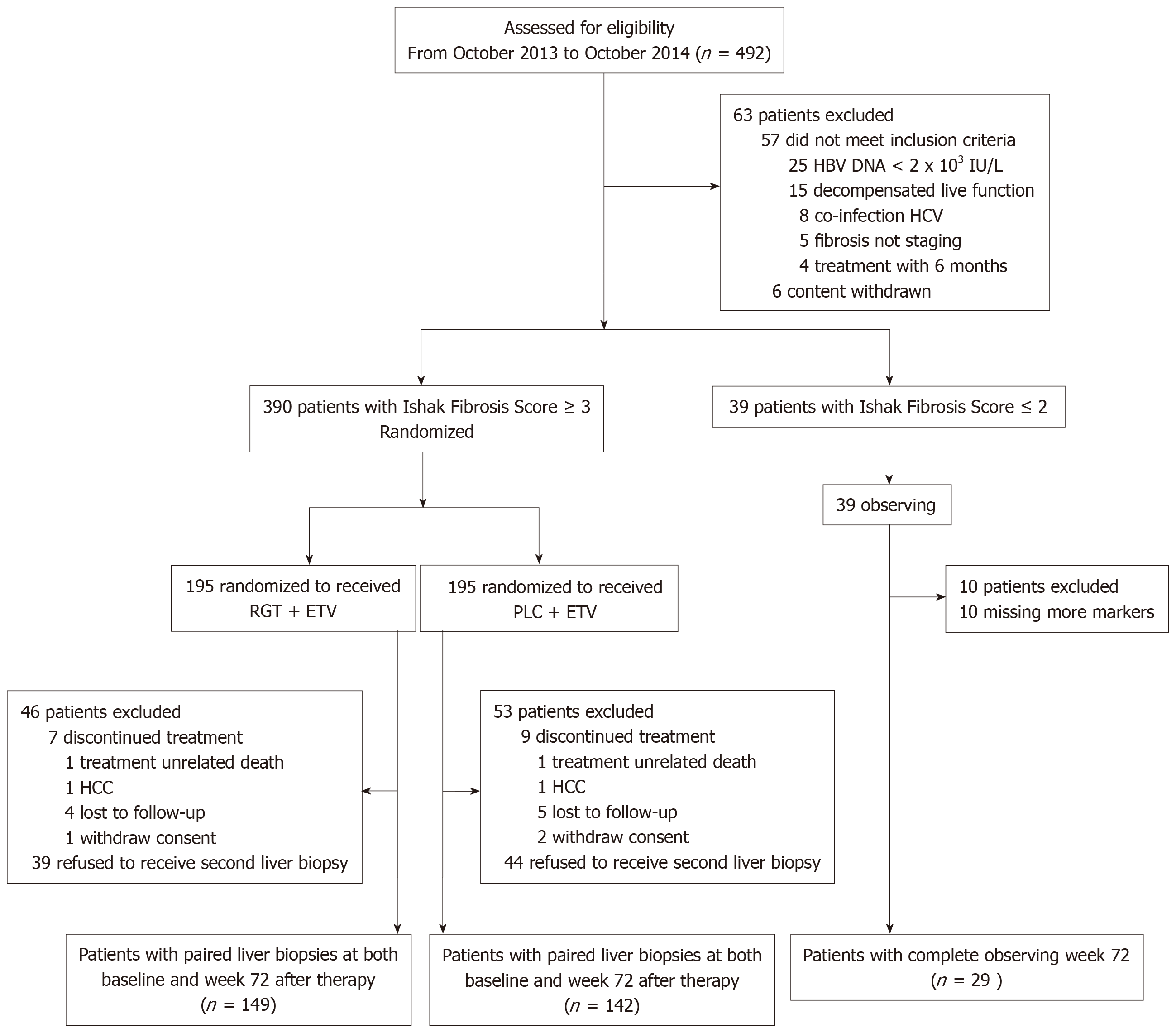
Figure 1 Flow chart of study population.
ETV: Entecavir; PLC: Placebo; RGT: Biejia-ruangan tablet; HCC: Hepatocellular carcinoma.
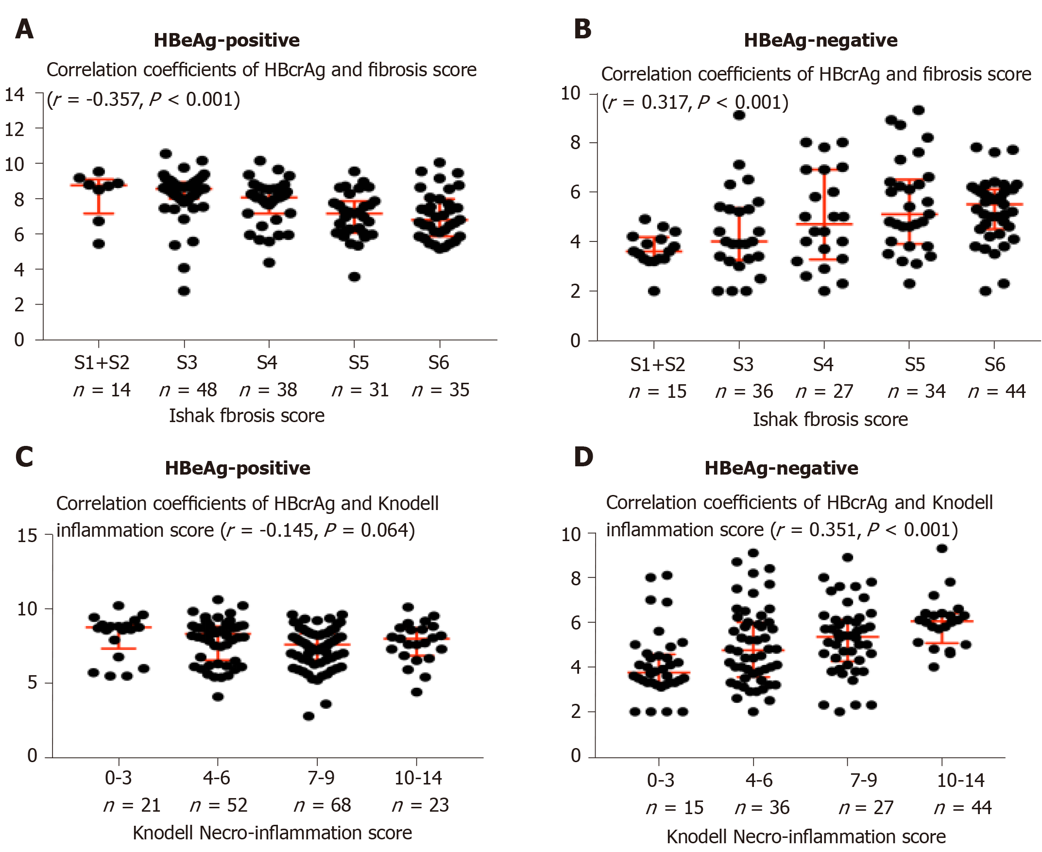
Figure 2 Correlations of hepatitis B core-related antigen with Ishak fibrosis score and Knodell necroflammation score in hepatitis B e antigen-positive (A and C) (n=164) and hepatitis B e antigen-negative (B and D) (n=156) patients.
Error bars, interquartile range; expression of measurement units: Hepatitis B core-related antigen, log10 kU/mL. HBcrAg: Hepatitis B core-related antigen.
Figure 3 Hepatitis B virus basal core promoter/precore/core mutations and serum concentration of hepatitis B core-related antigen.
A: Heat-map showing semi-supervised clustering of chronic hepatitis B (CHB) baseline hepatitis B core-related antigen (HBcrAg) level (X axis) between the basal core promoter/precure/core (BCP/PC/C) mutant (Y axis). According to the median of HBcrAg level, patients were divided into the low HBcrAg level (blue) and high HBcrAg level (yellow). Red color represents values higher than 2 standard deviations (SD) and blue color represents values lower than 2 SD from the mean value for each variable. B: Proportion of BCP/PC mutant in patients with low HBcrAg level (black bar) and high HBcrAg level (gray bar). C: HBcrAg level in patients with BCP/PC mutant (open box) and BCP/PC non-mutant (solid circle). D: Logarithmic reduction of HBcrAg in patients with BCP non-mutant (grey box) and BCP mutant (white box). BCP: Basal core promoter; PC: Precore; C: Core; CHB: Chronic hepatitis B; HBcrAg: Hepatitis B core-related antigen. Statistical significance is expressed as aP < 0.05, bP < 0.01 (P > 0.05 usually does not need to be denoted).
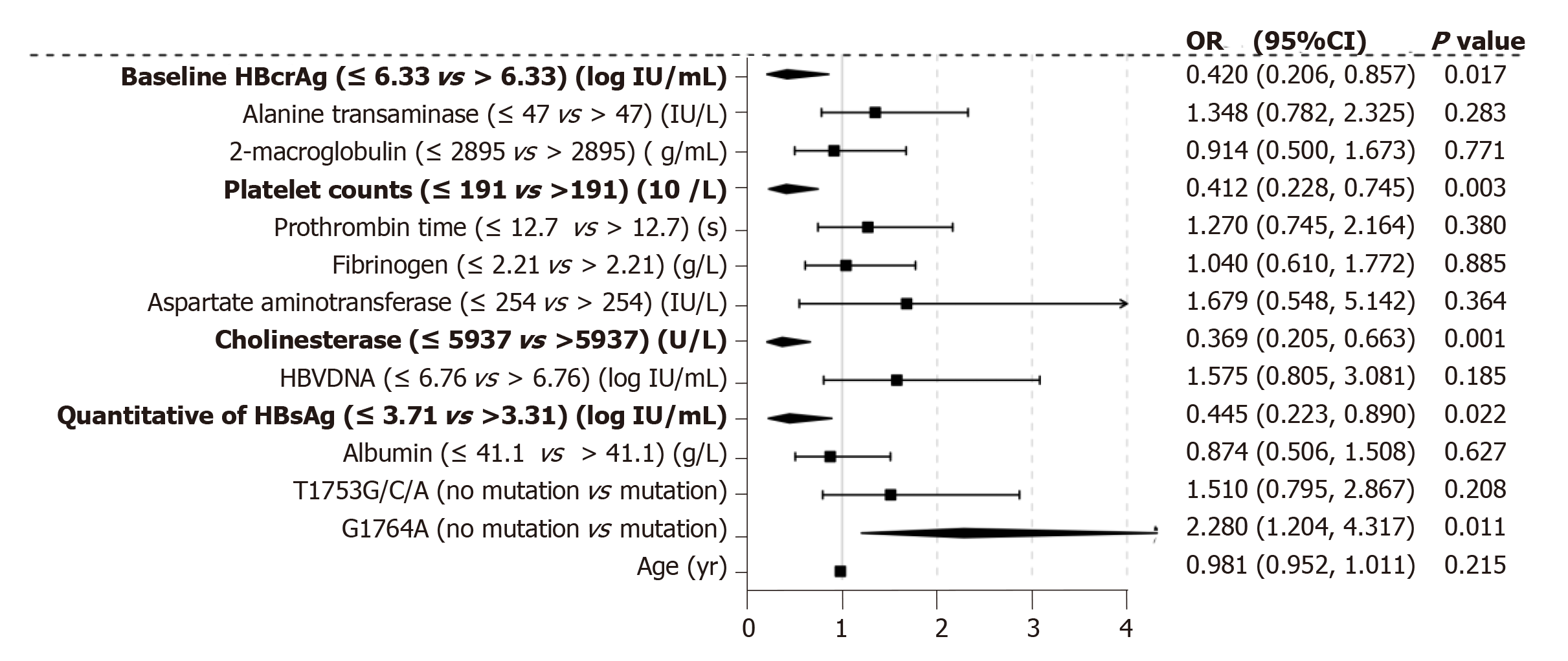
Figure 4 Variables associated with hepatic fibrosis.
Odds of Ishak fibrosis score 3-6 by multiple ordinal logistic regression for the whole patient. HBcrAg: Hepatitis B core-related antigen; OR: Odds rate; CI: Confidence interval.
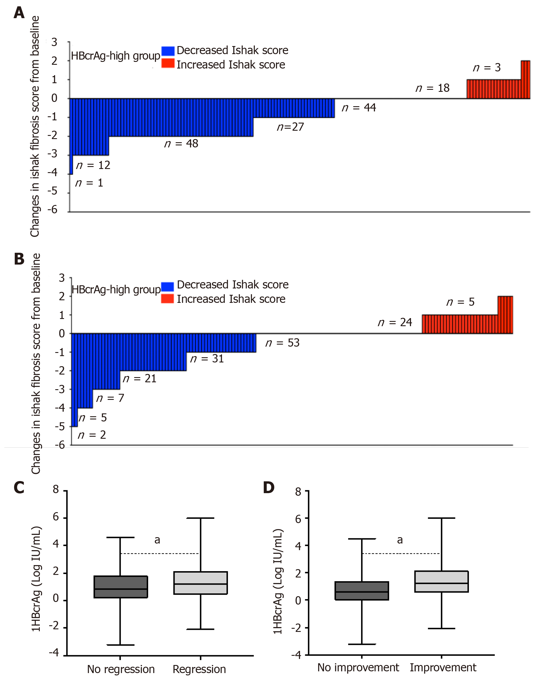
Figure 5 Hepatitis B core-related antigen kinetics and regression of liver fibrosis and histological improvement.
After entecavir therapy for 72 wk, regression of liver fibrosis in patients with low (A) and high (B) concentration of HBcrAg according to median baseline serum concentration of HBcrAg (6.33 log10 IU/mL, IQR: 4.80-8.18).Ishake fibrosis score decline is shown by blue, and increased is shown by red. C: 1HBcrAg as a median logarithmic reduction of HBcrAg from baseline in patients with regression of liver fibrosis (grey box) and non-regression (black box); and D: With the histological improvement (grey box) and non-improvement (black box). 1HBcrAg was defined as the baseline levels of HBcrAg minus week 72. Statistical significance is expressed as aP < 0.05, bP < 0.01 (P > 0.05 usually does not need to be denoted). HBcrAg: Hepatitis B core-related antigen; IQR: Interquartile range; OR: Odds rate.
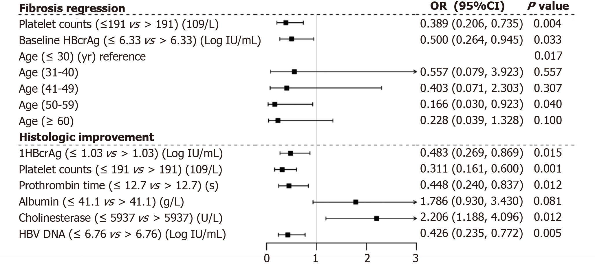
Figure 6 Variables associated with regression of liver fibrosis and histological improvement.
Odds of presence of the regression of liver fibrosis (upper) and histological improvement (down) by multiple ordinal logistic regression for the whole cohort of patients.
- Citation: Chang XJ, Sun C, Chen Y, Li XD, Yu ZJ, Dong Z, Bai WL, Wang XD, Li ZQ, Chen D, Du WJ, Liao H, Jiang QY, Sun LJ, Li YY, Zhang CH, Xu DP, Chen YP, Li Q, Yang YP. On-treatment monitoring of liver fibrosis with serum hepatitis B core-related antigen in chronic hepatitis B. World J Gastroenterol 2019; 25(32): 4764-4778
- URL: https://www.wjgnet.com/1007-9327/full/v25/i32/4764.htm
- DOI: https://dx.doi.org/10.3748/wjg.v25.i32.4764


