Copyright
©The Author(s) 2019.
World J Gastroenterol. Aug 28, 2019; 25(32): 4598-4613
Published online Aug 28, 2019. doi: 10.3748/wjg.v25.i32.4598
Published online Aug 28, 2019. doi: 10.3748/wjg.v25.i32.4598
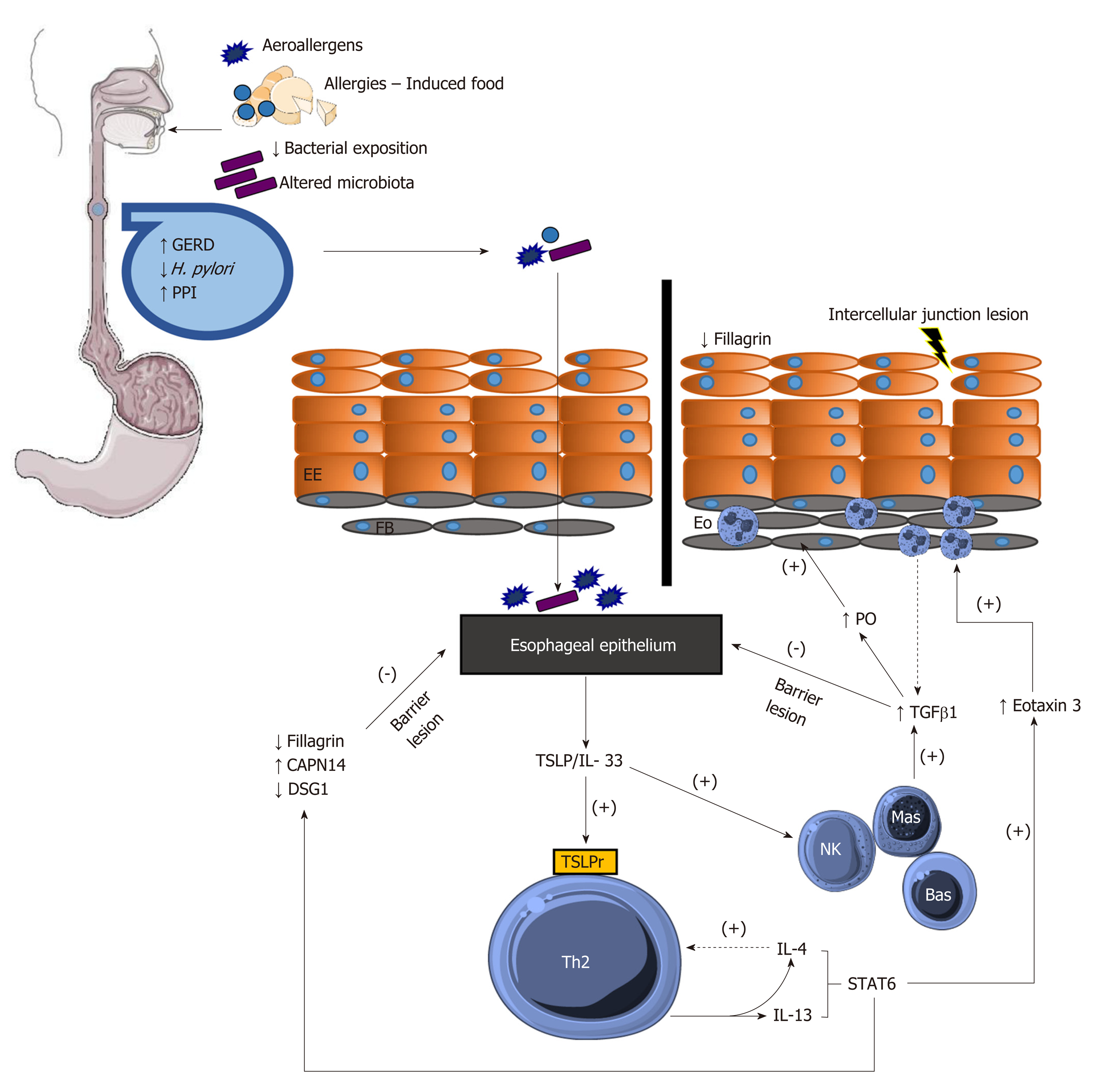
Figure 1 Pathophysiology of eosinophilic esophagitis.
TSLPr: TSLP receptor; IL-4r: IL-4 receptor; Eo: Eosinophils; Bas: Basophils; Mas: Mast cells; NK: Natural killer cells; PO: Periostin.
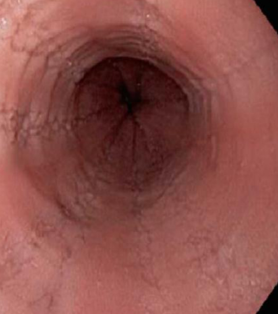
Figure 2 Longitudinal furrows in the esophagus.
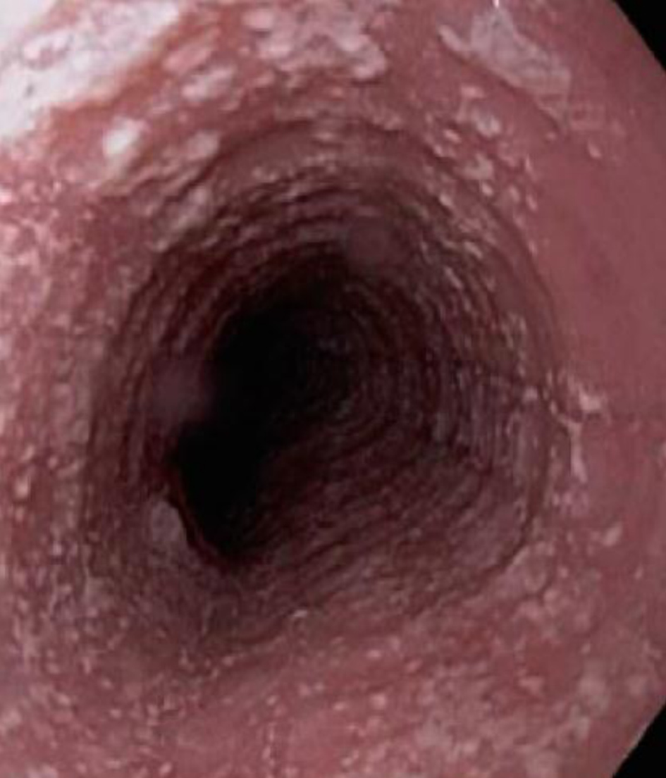
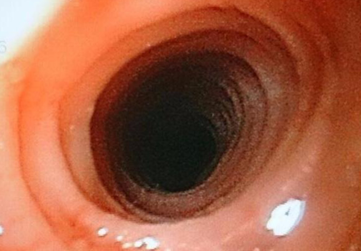
Figure 3 Whitish exudate in the esophagus and trachealized esophagus.
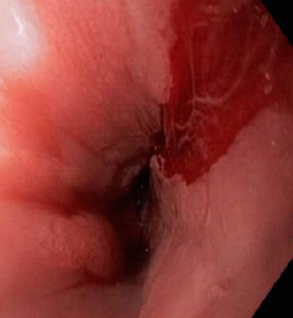
Figure 4 Superficial tear of the proximal esophageal mucosa.
Figure 5 Trachealized mucosa of the esophagus.
- Citation: Gómez-Aldana A, Jaramillo-Santos M, Delgado A, Jaramillo C, Lúquez-Mindiola A. Eosinophilic esophagitis: Current concepts in diagnosis and treatment. World J Gastroenterol 2019; 25(32): 4598-4613
- URL: https://www.wjgnet.com/1007-9327/full/v25/i32/4598.htm
- DOI: https://dx.doi.org/10.3748/wjg.v25.i32.4598


