Copyright
©The Author(s) 2019.
World J Gastroenterol. Aug 21, 2019; 25(31): 4320-4342
Published online Aug 21, 2019. doi: 10.3748/wjg.v25.i31.4320
Published online Aug 21, 2019. doi: 10.3748/wjg.v25.i31.4320
Figure 1 History of the ileal pouch.
A: The Kock continent ileostomy; B: Common pouch configurations used in ileal pouches.
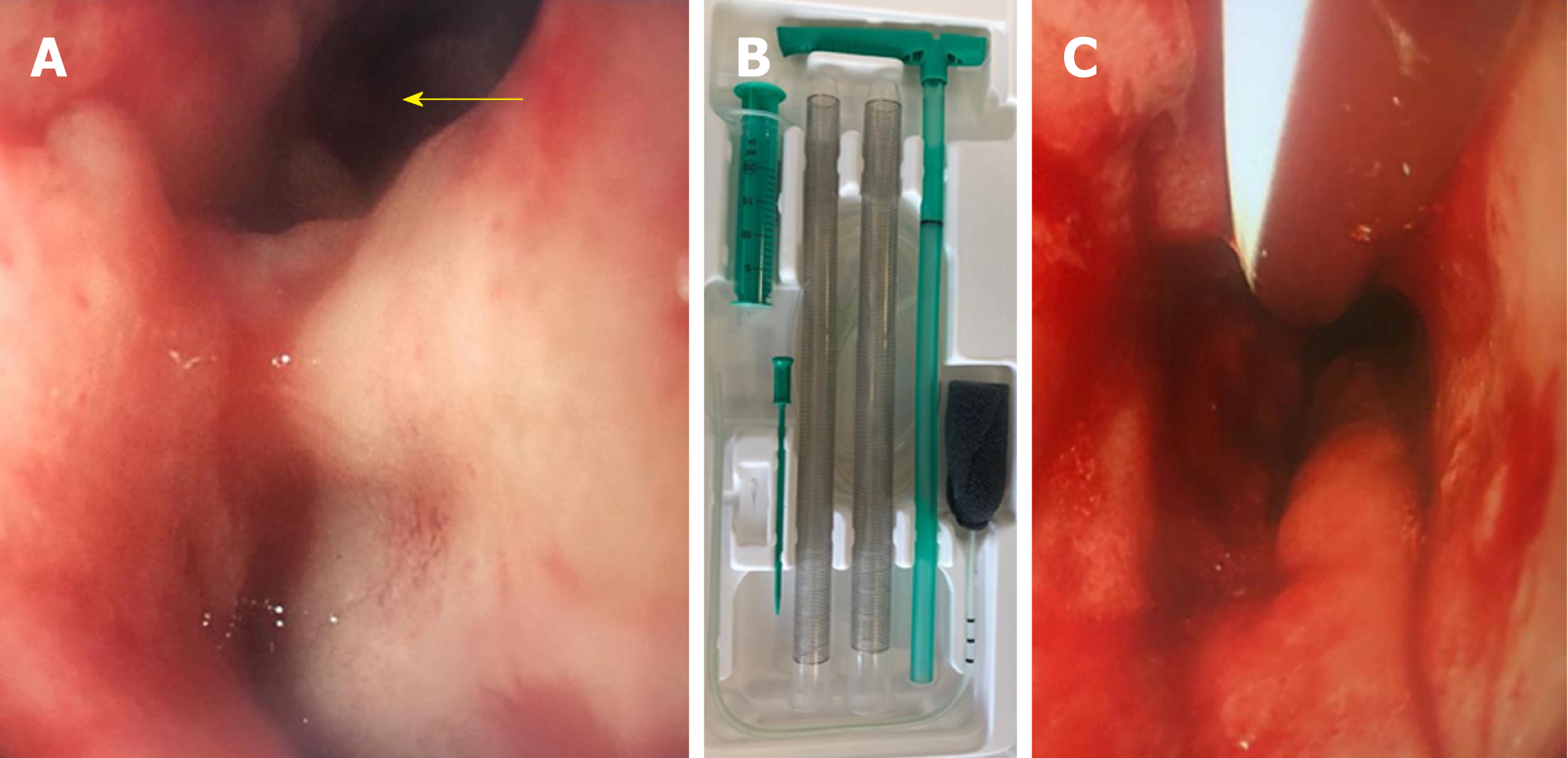
Figure 2 Endo-cavitational vacuum therapy (Endo-SPONGE®).
A: Endoscopic view of a pouch with a cavity (arrow) at the anastomosis secondary to anastomotic leak. The true lumen is at the inferior aspect; B: Contents of the Endo-SPONGE® kit (B Braun Medical Ltd); C: The foam of the Endo-SPONGE® is passed into the cavity and negative pressure applied to collapse the cavity.
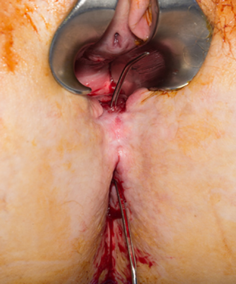
Figure 3 Examination under anaesthesia performed for a female patient who presented with perineal sepsis and abnormal per vaginal discharge following ileal pouch anal anastomosis.
A pouch-vaginal fistula was identified and confirmed with a probe.
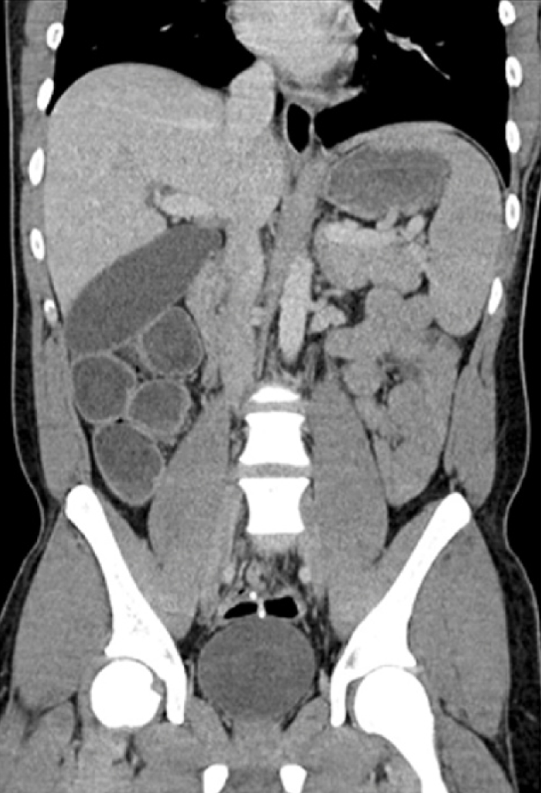
Figure 4 A coronal computed tomography image of a patient who presented with early small bowel obstruction following closure of ileostomy (6 mo post pouch creation).
Computed tomography and operative findings confirmed small bowel obstruction secondary to a 360° twist at the level of the anastomosis.
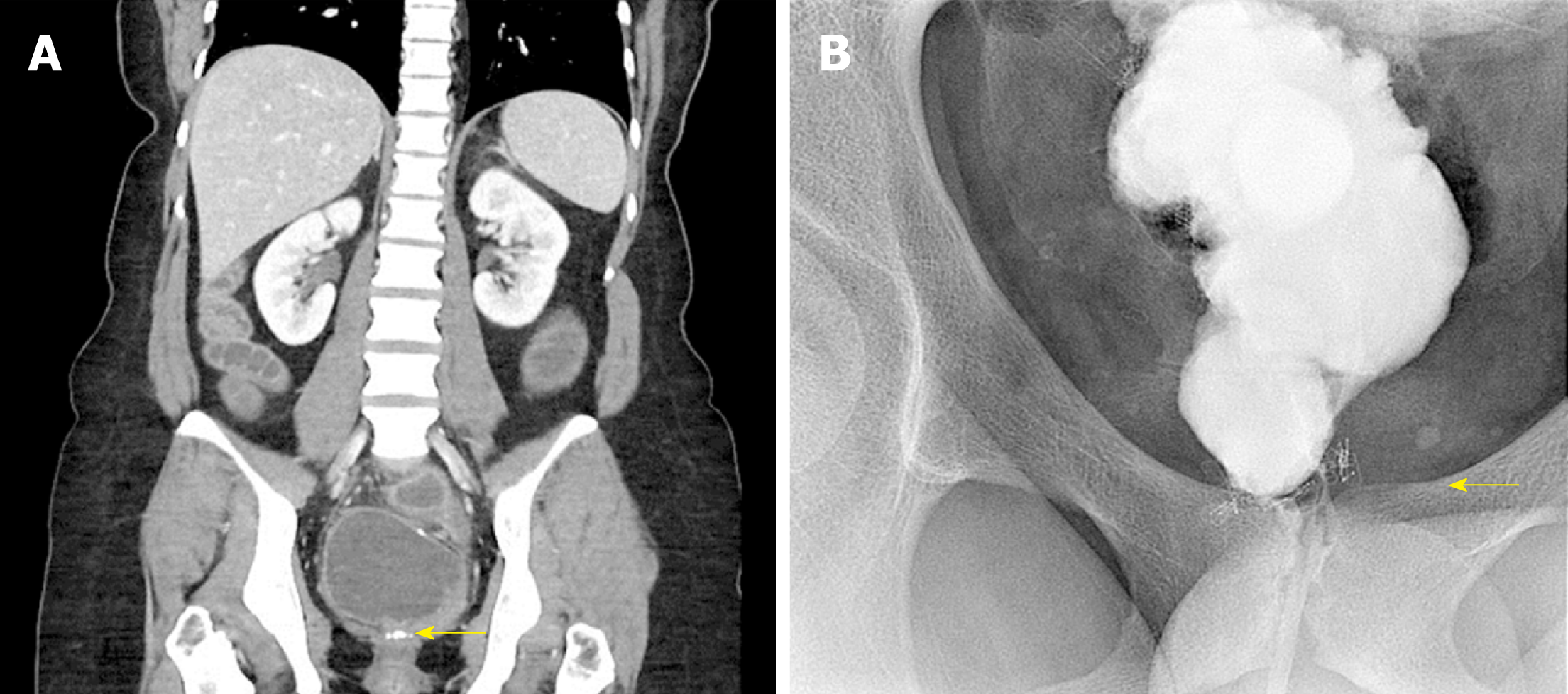
Figure 5 Obstruction to pouch outflow usually occurs at the level of the anastomosis.
A: A coronal computed tomography image demonstrating a pouch outlet stricture. A stricture at the level of the anastomosis (arrow) caused a dilated pouch that could not empty without intubation; B: A pouchogram of the same patient confirmed an anastomotic stricture that eventually yielded to serial Hegar dilations.
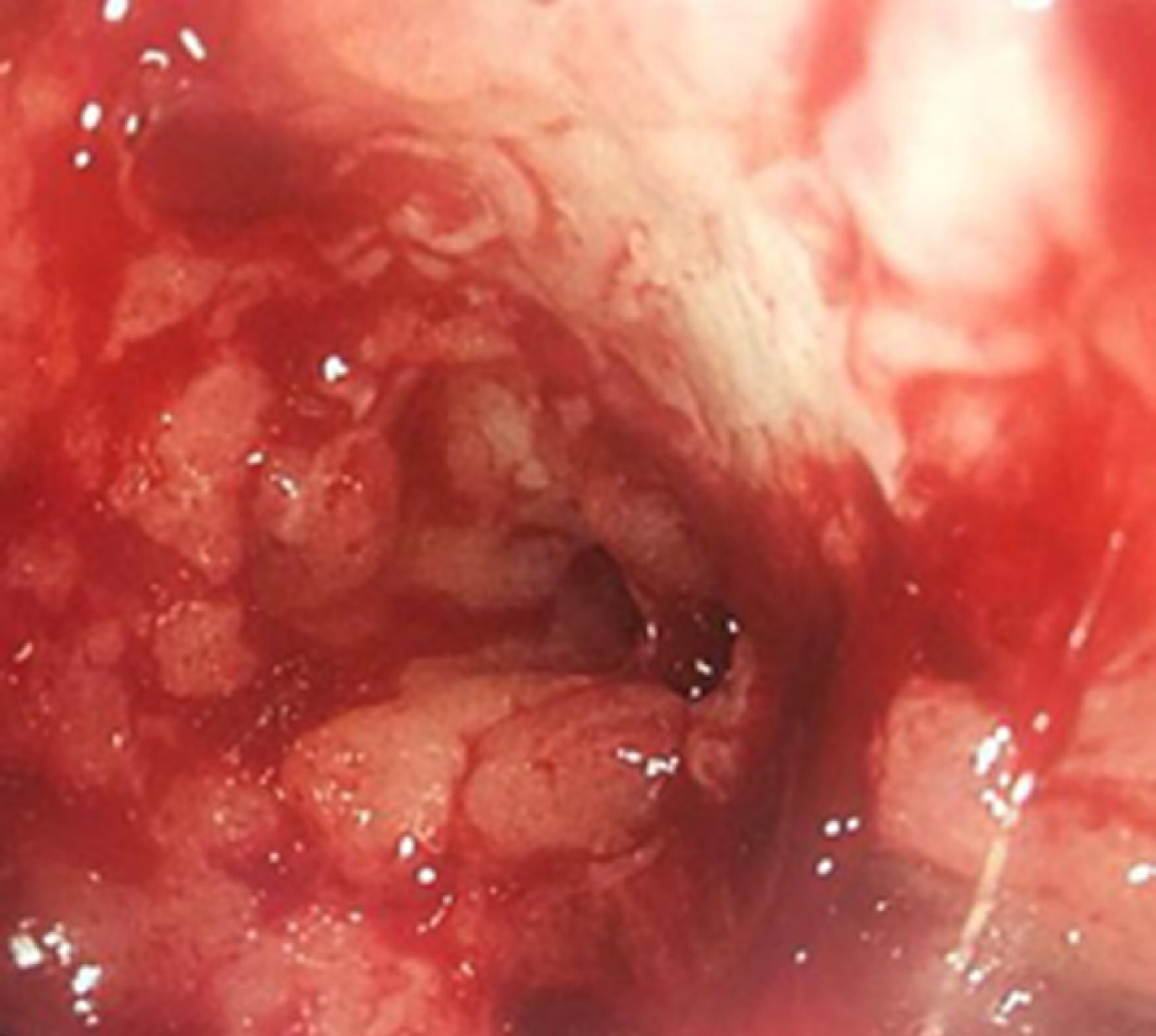
Figure 6 Endoscopic view of pouchitis (Pouch Disease Activity Index endoscopic sub-score 4).
Figure 7 Pouch adenoma.
A: Magnetic resonance image and endoscopic view of a pedunculated pouch polyp (arrow) arising in the mid pouch that was completely excised endoscopically; B: A serrated, near circumferential, lesion (arrow) that required formal pouch excision. Both pouch lesions were confirmed histologically to be pouch adenomas.
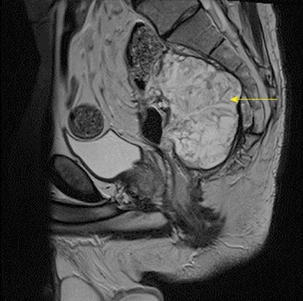
Figure 8 A magnetic resonance image of a pouch with a large exophytic lesion (arrow) arising from its posterior aspect.
Biopsies and formal histopathology confirmed this to be a pouch carcinoma.
- Citation: Ng KS, Gonsalves SJ, Sagar PM. Ileal-anal pouches: A review of its history, indications, and complications. World J Gastroenterol 2019; 25(31): 4320-4342
- URL: https://www.wjgnet.com/1007-9327/full/v25/i31/4320.htm
- DOI: https://dx.doi.org/10.3748/wjg.v25.i31.4320


