Copyright
©The Author(s) 2019.
World J Gastroenterol. Aug 14, 2019; 25(30): 4061-4073
Published online Aug 14, 2019. doi: 10.3748/wjg.v25.i30.4061
Published online Aug 14, 2019. doi: 10.3748/wjg.v25.i30.4061
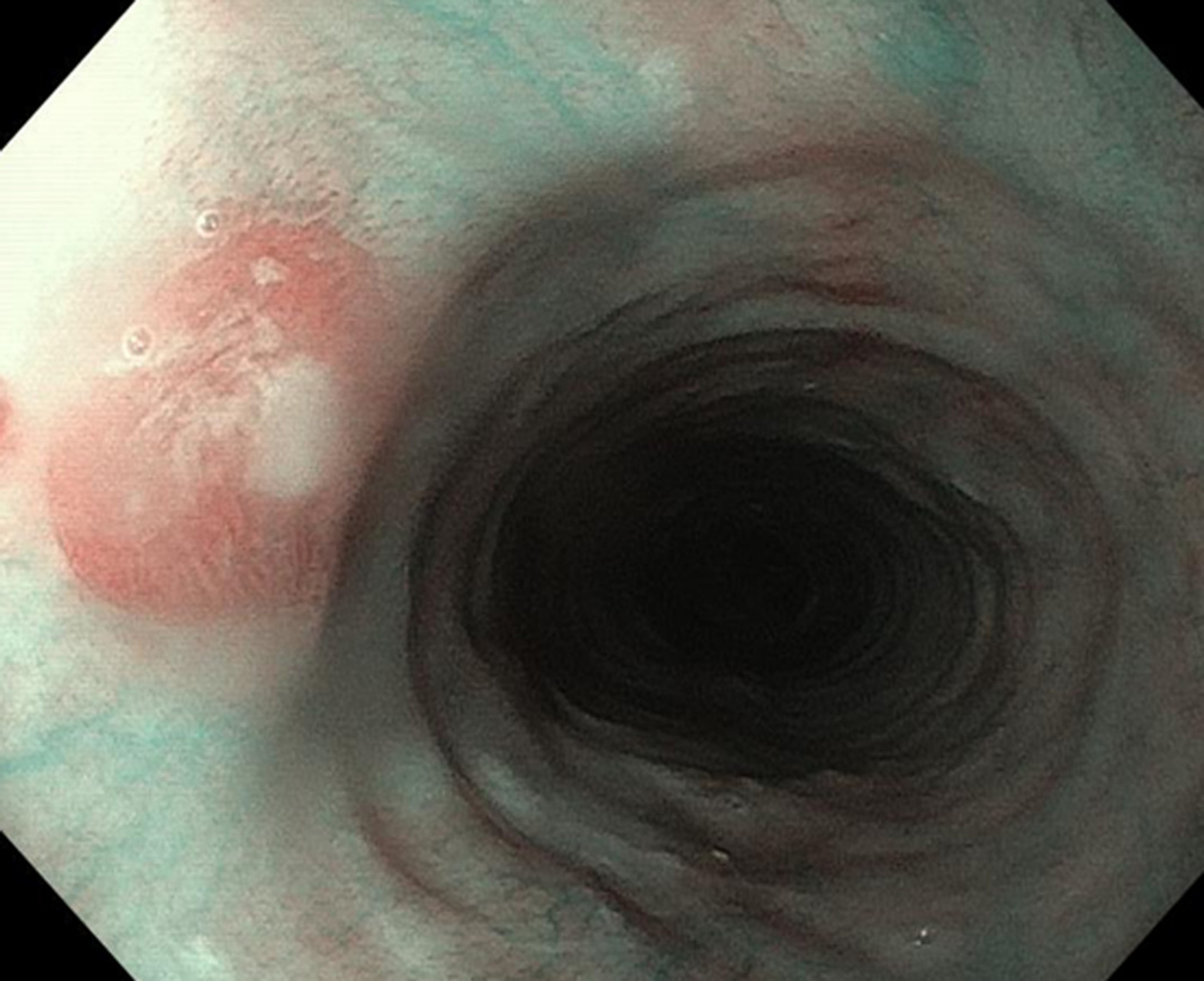
Figure 1 A discreet area of flat salmon pink mucosa with mucus on surface typically found in the proximal esophagus of a 45-year-old anxious woman with globus sensation.
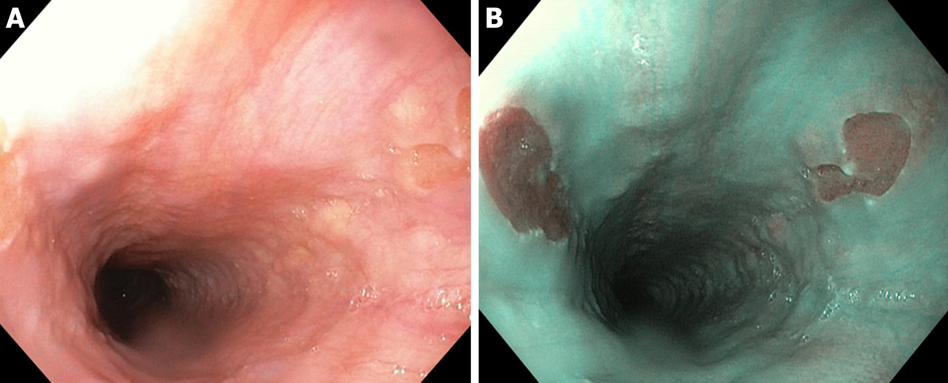
Figure 2 Double mirror flat inlet patches in (A) white light endoscopy vs (B) optical chromoendoscopy (narrow band imaging), in a middle age woman with Helicobacter pylori-associated gastritis and globus sensation ameliorated after the eradication therapy.
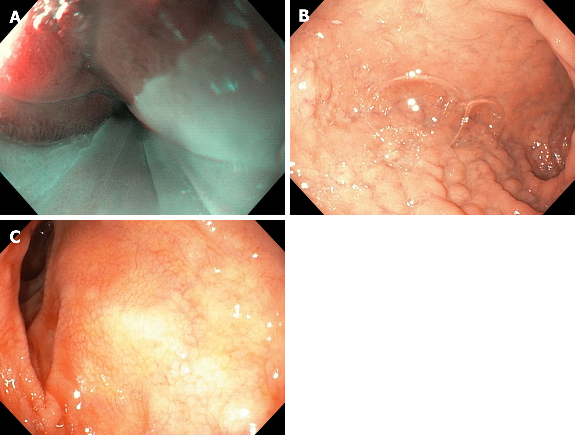
Figure 3 Concomitant findings of inlet patches in patients with inflammatory bowel disease, celiac disease, neurofibromatosis or blue rubber bleb nevus syndrome are likely to be incidental.
A: Large inlet patch from 16 to 20 cm from the incisors in a young woman with globus sensation and concomitant celiac disease. The patient underwent an upper endoscopy because of persistent iron deficiency anemia. B: Nodular appearance of duodenal mucosa and C: flattened villi in the same patient.
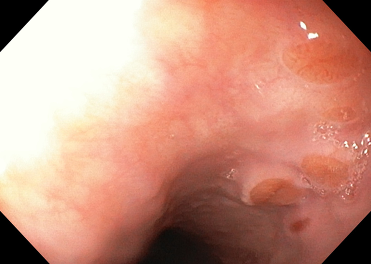
Figure 4 Multiple small focal areas round in shape of gastric tissue, one of them slightly raised, noted in the right lateral field, 10 cm from the incisors, in a young man presenting for unexplained upper dysphagia.
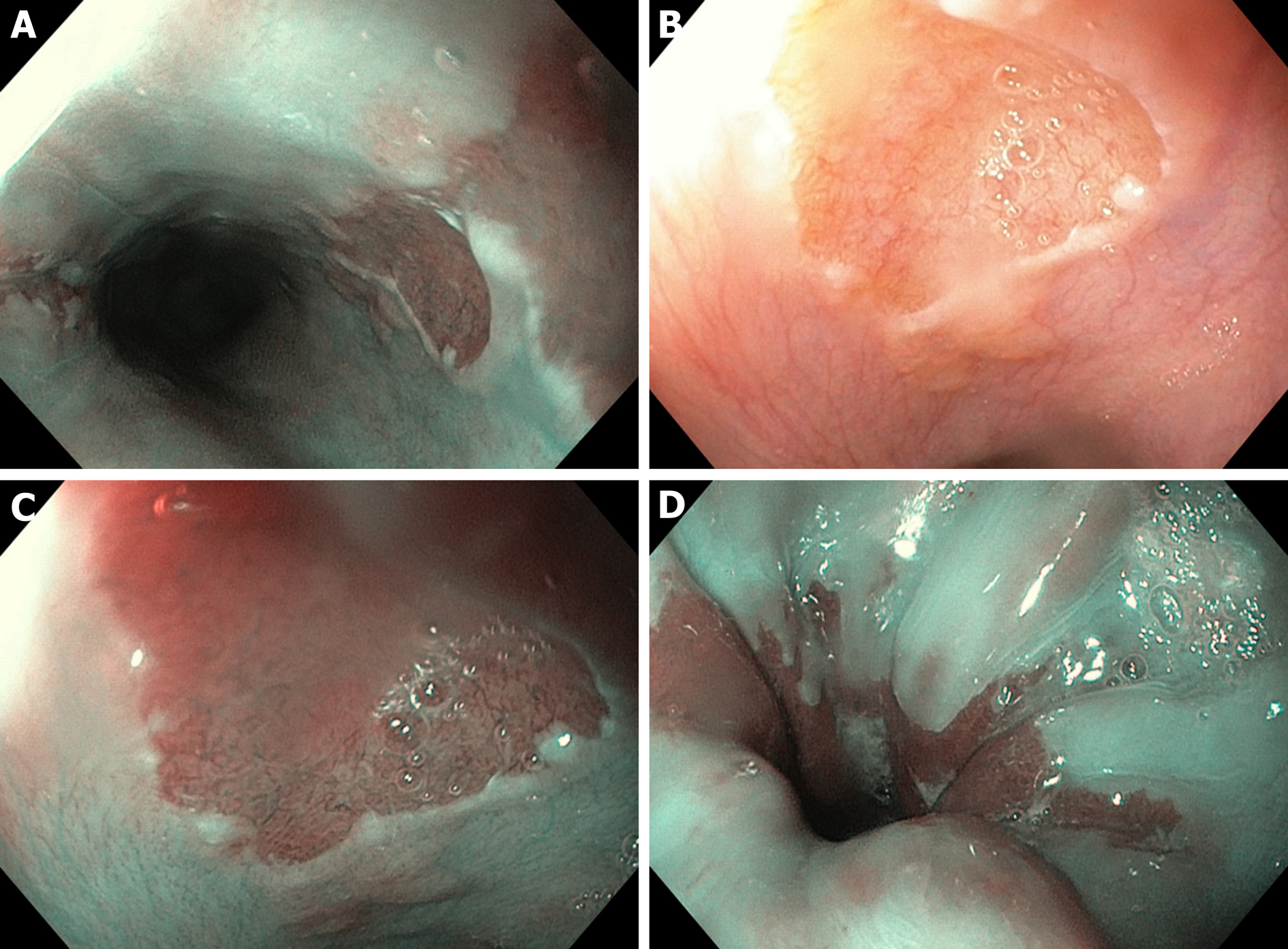
Figure 5 A: Three areas of cervical inlet patches, with kissing distribution, in a middle age women with uterine cancer history, presenting for reflux complaints and globus sensation.
Detailed image in (B) white light endoscopy and (C) narrow band imaging. D: Irregular Z line in the same patient suggesting concomitant gastroesophageal reflux disease.
- Citation: Ciocalteu A, Popa P, Ionescu M, Gheonea DI. Issues and controversies in esophageal inlet patch. World J Gastroenterol 2019; 25(30): 4061-4073
- URL: https://www.wjgnet.com/1007-9327/full/v25/i30/4061.htm
- DOI: https://dx.doi.org/10.3748/wjg.v25.i30.4061


