Copyright
©The Author(s) 2019.
World J Gastroenterol. Jul 14, 2019; 25(26): 3370-3379
Published online Jul 14, 2019. doi: 10.3748/wjg.v25.i26.3370
Published online Jul 14, 2019. doi: 10.3748/wjg.v25.i26.3370
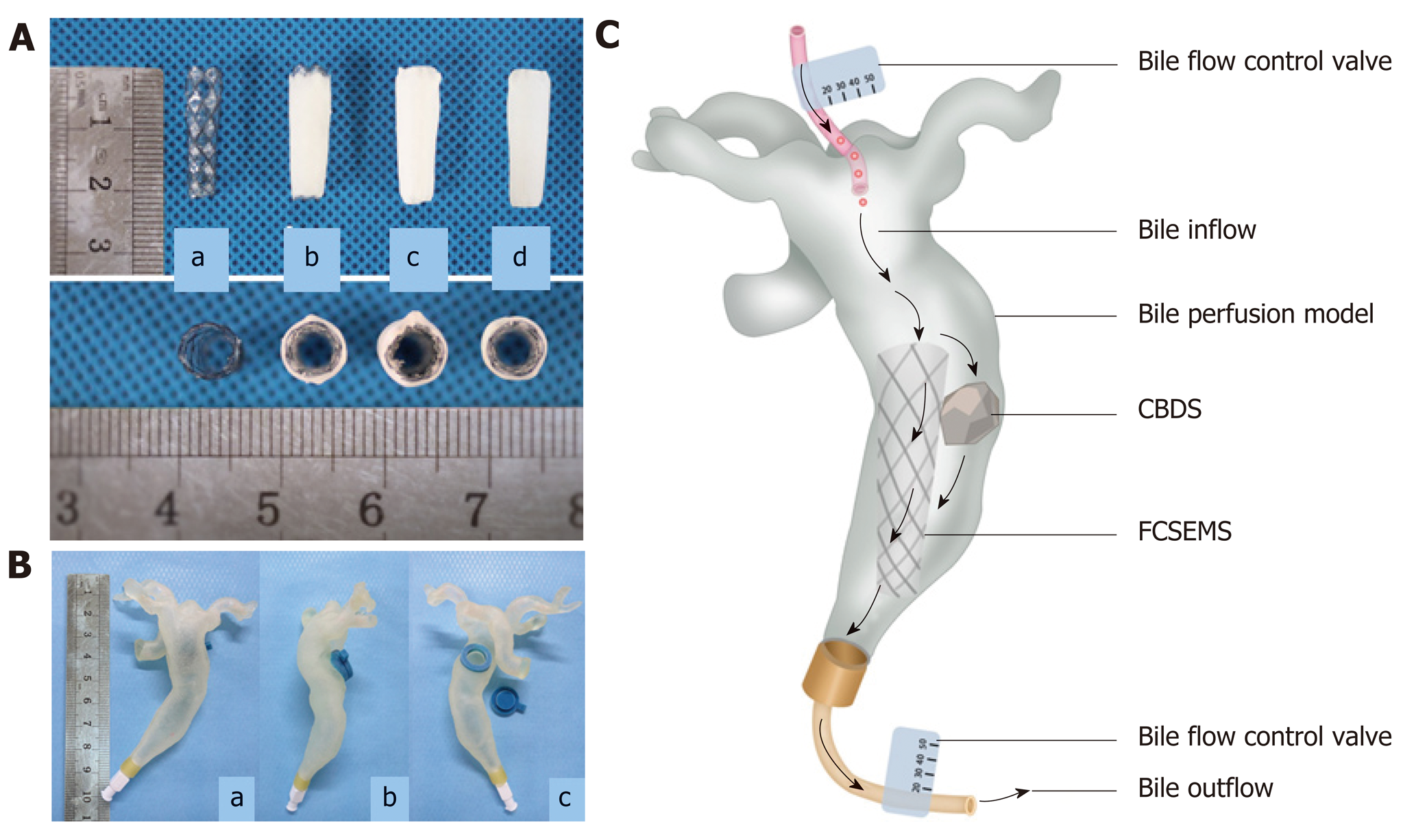
Figure 1 Pictures of stents and common bile duct model.
A: Picture of covered nitinol stent and drug-eluting stents manufactured by dip coating, coaxial electrospinning, and dip coating combined with electrospinning (a: Covered nitinol stent without drug load; b: Drug-eluting stent manufactured by dip coating; c: Drug-eluting stent manufactured by coaxial electrospinning; d: Drug-eluting stent manufactured by dip coating combined with electrospinning); B: In vitro common bile duct (CBD) model manufactured by 3D printing technology (a: Front view; b: Lateral view; c: Back view, one stent and one stone can be put inside the model through the hole on the back); C: Picture illustrating dissolution of biliary stone in flowing bile in bile perfusion model. One stent and one CBD stone (mass in 100 mg) were put into the CBD model together and a total of 1000 mL human bile was perfused into each CBD model every day, that is, 1000 mL human bile will flow through the stent and stone every day. In this way, we get to know whether our stent can exert the stone-dissolving effect in flowing bile. CBD: Common bile duct; CBDS: Common bile duct stone.
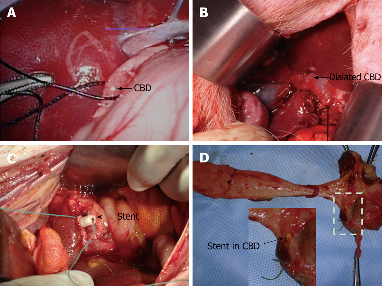
Figure 2 Key steps to put stent into porcine common bile duct.
A: The porcine common bile duct (CBD) was partially ligated via laparoscopy; B: The porcine CBD was dilated after one-week ligation; C: Naked fully covered self-expanding metal stent or Stent III was put into the porcine CBD through choledochotomy. To avoid stent migration, stents were sewed on the CBD walls with special care when closing the CBD and the ligation was removed thereafter; D: After the animals were sacrificed, all the stents were still in the bile duct without migration. CBD: Common bile duct.
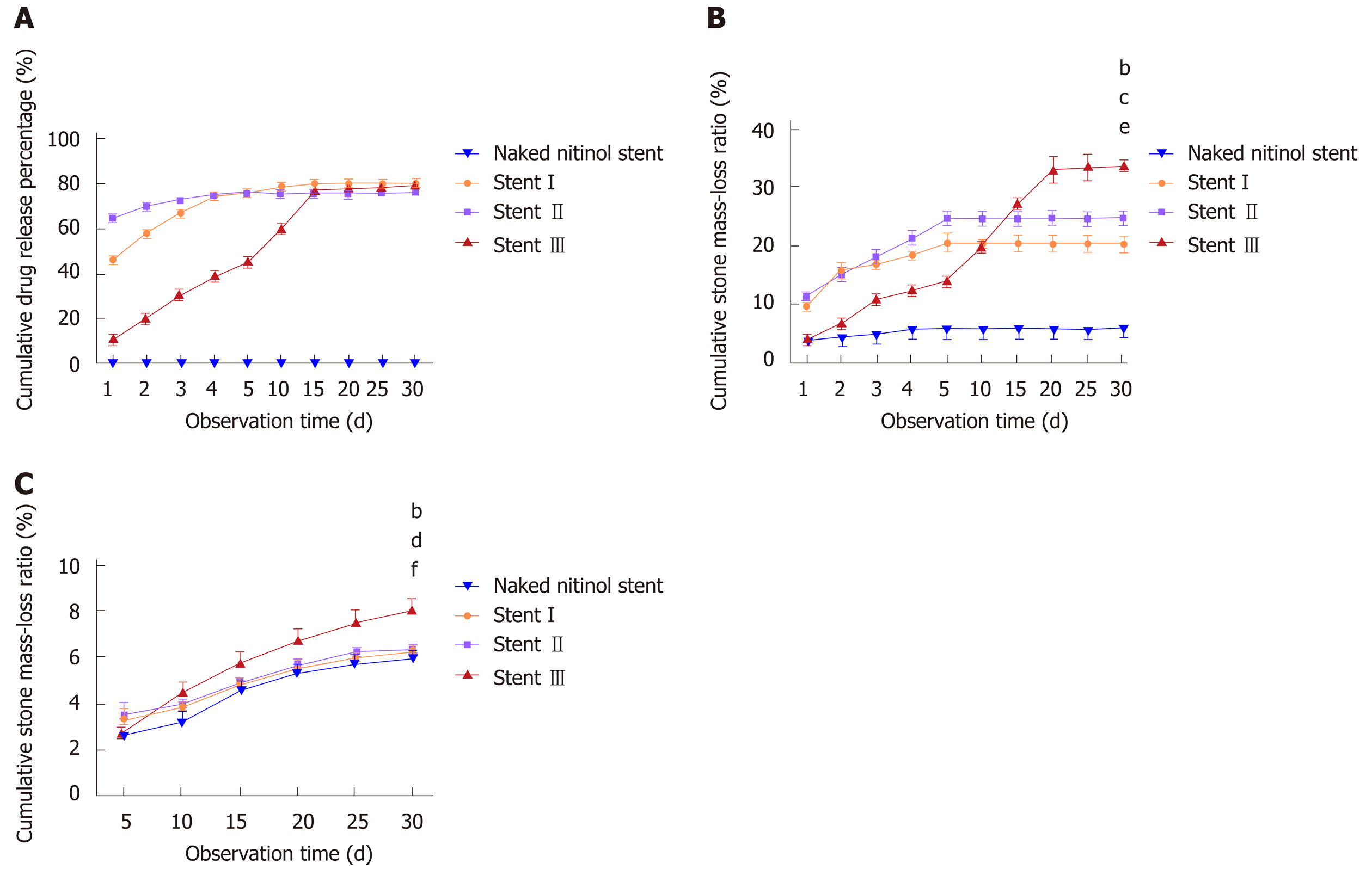
Figure 3 Drug release behavior and stone-dissolving efficacy of different stents.
A: Drug release curves of three types of drug-eluting stents and stents without drug load; B: Stone mass loss curves of three types of drug-eluting stents and stents without drug load in still buffer; C: Stone mass loss curves of three types of drug-eluting stents and stents without drug load in flowing bile. Naked fully covered self-expanding metal stent vs Stent III, aP < 0.05, bP < 0.01; Stent I vs Stent III, cP < 0.05, dP < 0.01; Stent II vs Stent III, eP < 0.05, fP < 0.01.
Figure 4 Pictures of hematoxylin and eosin staining of gallbladder, common bile duct, duodenum, liver, and kidney tissues after 30-d placement of naked fully covered self-expanding metal stent or Stent III in the porcine common bile duct (100-fold magnification).
- Citation: Huang C, Cai XB, Guo LL, Qi XS, Gao Q, Wan XJ. Drug-eluting fully covered self-expanding metal stent for dissolution of bile duct stones in vitro. World J Gastroenterol 2019; 25(26): 3370-3379
- URL: https://www.wjgnet.com/1007-9327/full/v25/i26/3370.htm
- DOI: https://dx.doi.org/10.3748/wjg.v25.i26.3370


