Copyright
©The Author(s) 2019.
World J Gastroenterol. May 28, 2019; 25(20): 2473-2488
Published online May 28, 2019. doi: 10.3748/wjg.v25.i20.2473
Published online May 28, 2019. doi: 10.3748/wjg.v25.i20.2473
Figure 1 Schematic diagram of the Th17 cell differentiation pathway.
APC: Antigen-presenting cells; TGF-β: transforming growth factor β; IL: Interleukin; IL-23R: Interleukin-23 receptor 1.
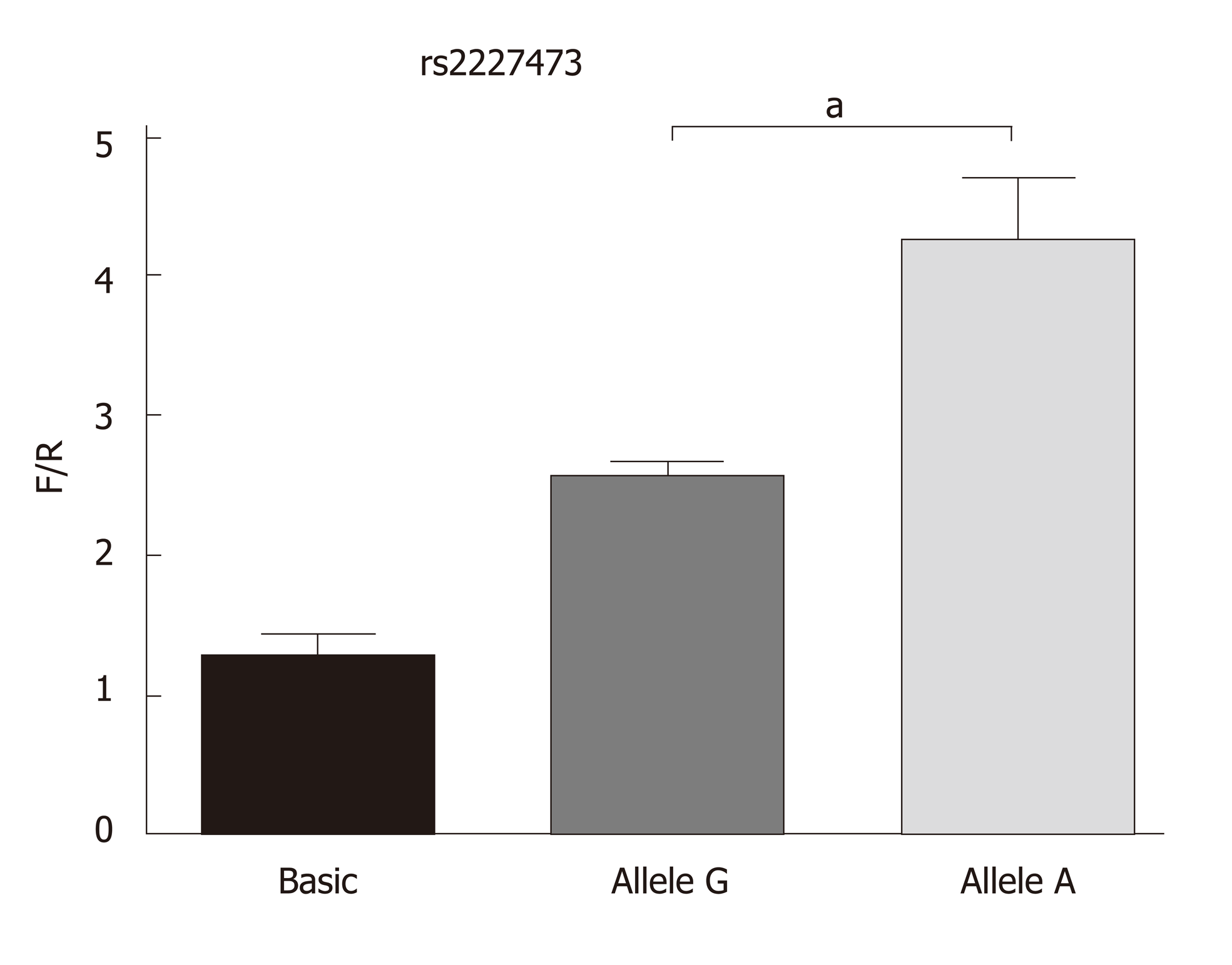
Figure 2 Fluorescence detected in cells transfected with the recombinant or empty vector.
Allele G and allele A indicate the fluorescence results of cells transfected with the pGL3-rs2227473_G and pGL3-rs2227473_A plasmids, respectively. F/R: Firefly/renilla luciferase values; aP < 0.05.
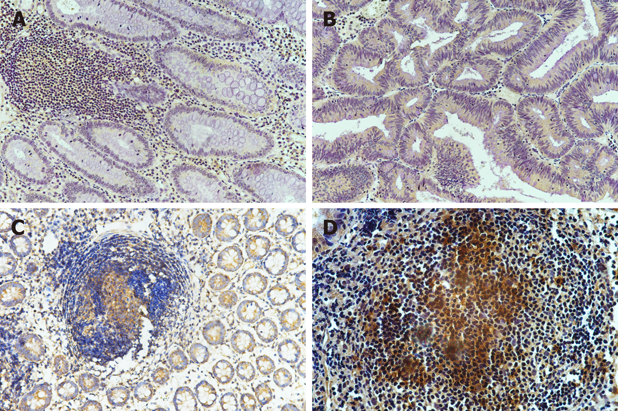
Figure 3 Immunohistochemical staining for interleukin-22 receptor 1 in intestinal biopsy tissues.
A: Colon polyps; B: Colon cancer; C: Crohn's disease; D: Intestinal tuberculosis. Magnification, 100×.
Figure 4 In typical caseous necrotic granulomas, Langhans giant cells with high interleukin-22 receptor 1 expression are present at the edge of granulomas.
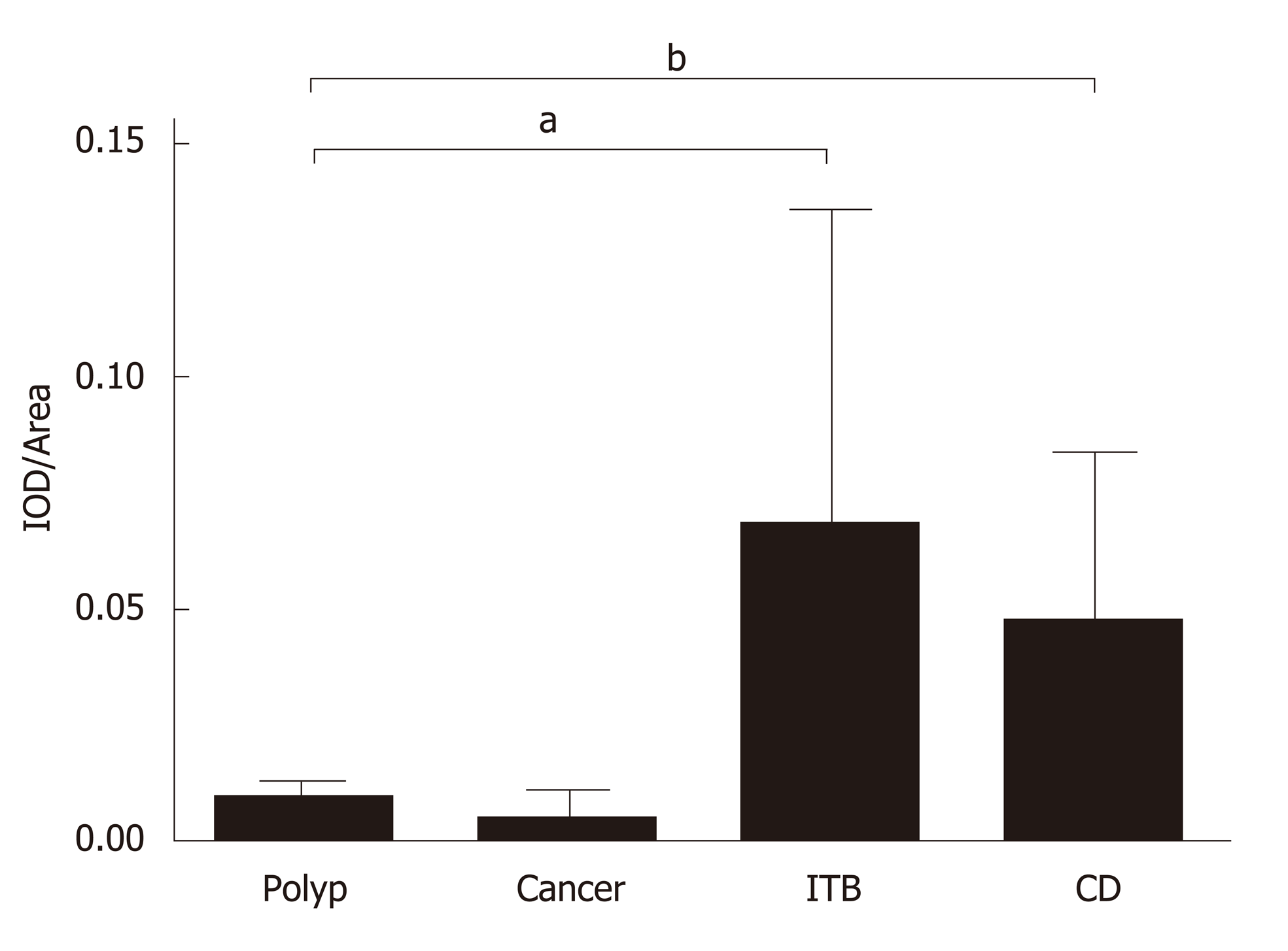
Figure 5 Total integrated optical density/total area of immunohistochemical images.
IOD: Integrated optical density; CD: Crohn's disease; ITB: Intestinal tuberculosis; aP < 0.05, bP < 0.01.
- Citation: Yu ZQ, Wang WF, Dai YC, Chen XC, Chen JY. Interleukin-22 receptor 1 is expressed in multinucleated giant cells: A study on intestinal tuberculosis and Crohn's disease. World J Gastroenterol 2019; 25(20): 2473-2488
- URL: https://www.wjgnet.com/1007-9327/full/v25/i20/2473.htm
- DOI: https://dx.doi.org/10.3748/wjg.v25.i20.2473


