Copyright
©The Author(s) 2019.
World J Gastroenterol. Jan 14, 2019; 25(2): 205-219
Published online Jan 14, 2019. doi: 10.3748/wjg.v25.i2.205
Published online Jan 14, 2019. doi: 10.3748/wjg.v25.i2.205
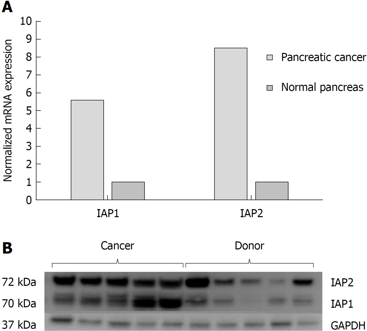
Figure 1 Pancreatic cancer specimens displayed increased Inhibitors of apoptosis proteins expression analysis in cancer tissues.
mRNA and protein expression of inhibitors of apoptosis proteins in normal tissues (n = 9) and pancreatic cancer (n = 61) were evaluated by A: quantitative reverse transcription polymerase chain reaction; and B: western blot analysis. IAP1: Inhibitor of apoptosis protein 1; IAP2: Inhibitor of apoptosis protein 2; GAPDH: Glyceraldehyde-3-phosphate dehydrogenase.
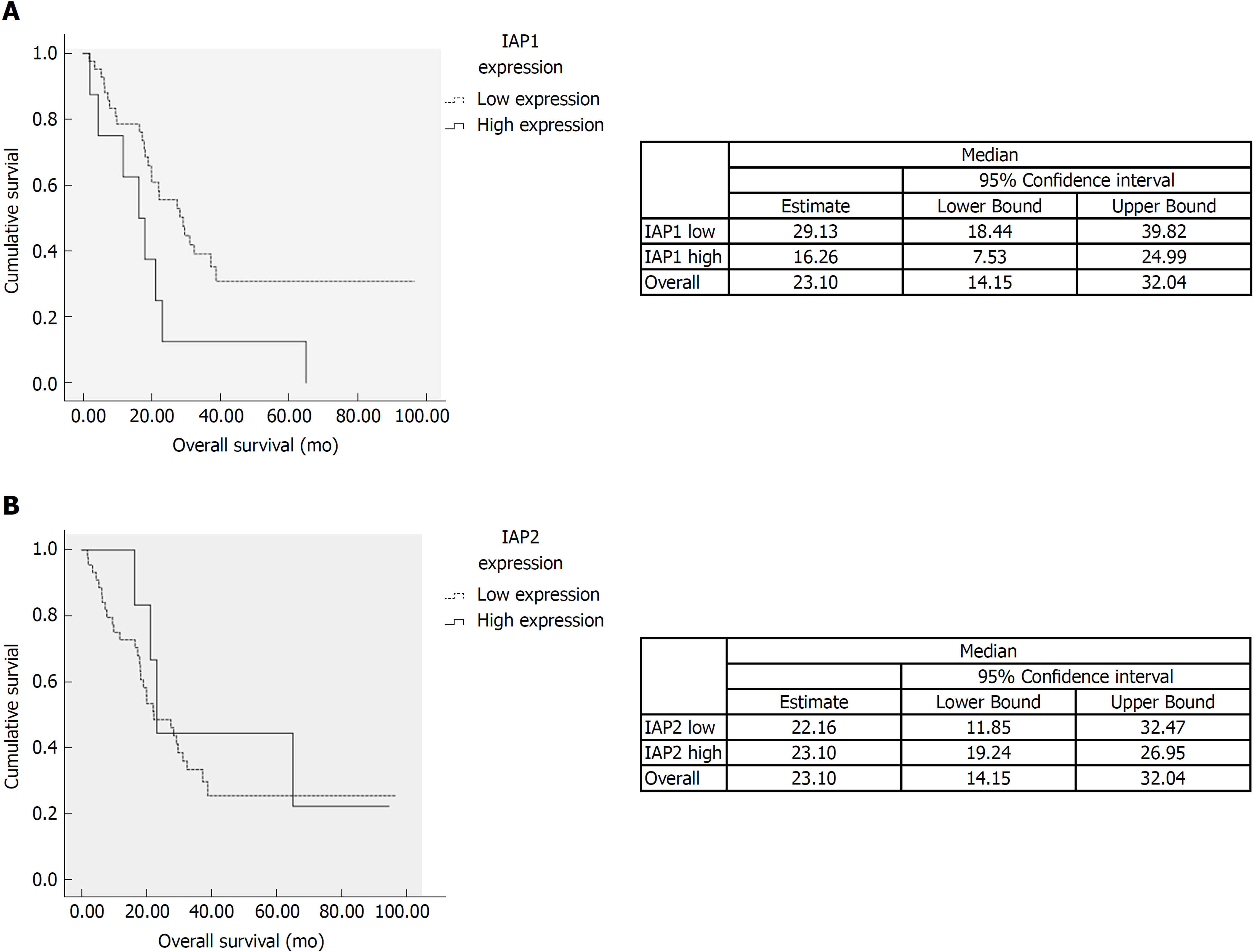
Figure 2 Survival analysis.
A: The survival time of patients with the low expression of inhibitor of apoptosis protein 1 tended to be longer than those with high expression (P < 0.05). B: There was no difference of survival among high and low inhibitor of apoptosis protein 2 (IAP2) mRNA expression. IAP1: Inhibitor of apoptosis protein 1; IAP2: Inhibitor of apoptosis protein 2.
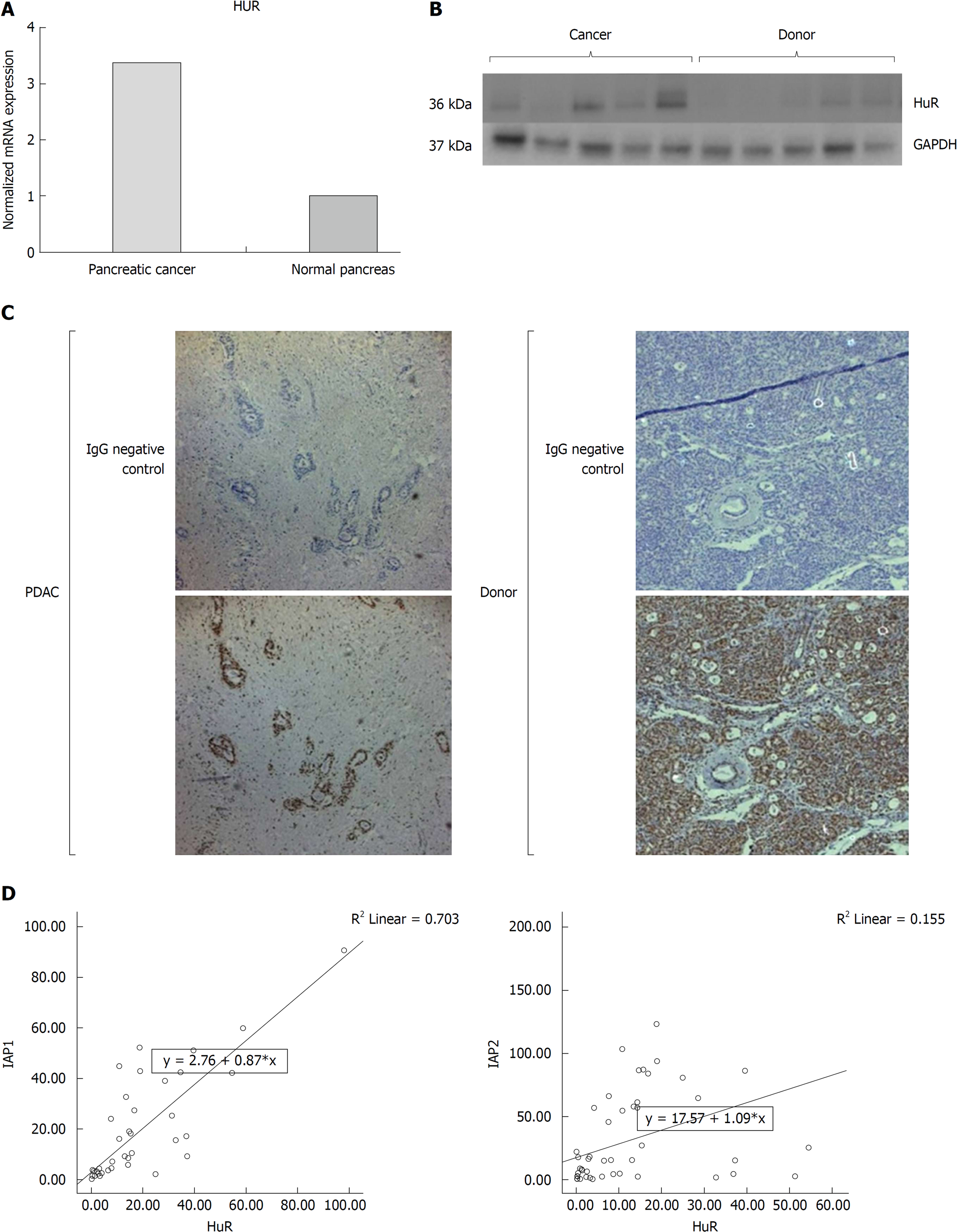
Figure 3 Pancreatic cancer specimens displayed increased human antigen R expression analysis in cancer tissues.
mRNA and protein expression of human antigen R (HuR) were upregulated by A: quantitative reverse transcription polymerase chain reaction; and B: western blot analysis. C: Immunohistochemistry showed that HuR was mainly positive in the ductal cancer cell’s nucleus and less in cytoplasm. D: Expressions of inhibitors of apoptosis proteins were correlated with HuR expression. IAP1: Inhibitor of apoptosis protein 1; IAP2: Inhibitor of apoptosis protein 2; HuR: Human antigen R; GAPDH: Glyceraldehyde-3-phosphate dehydrogenase; PDAC: Pancreatic ductal adenocarcinoma.
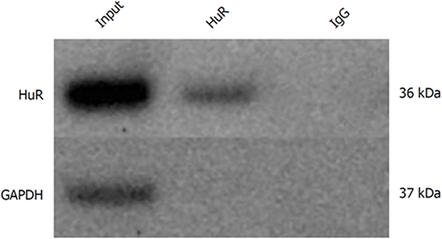
Figure 4 Human antigen R protein binds to inhibitors of apoptosis proteins mRNA in pancreatic cancer cells.
Human antigen R (HuR) and Glyceraldehyde-3-phosphate dehydrogenase protein levels in whole cell lysates (input), magnetic beads with anti-HuR antibody and protein precipitates (HuR), and magnetic beads with anti-IgG and protein precipitates (IgG). HuR: Human antigen R; GAPDH: Glyceraldehyde-3-phosphate dehydrogenase.
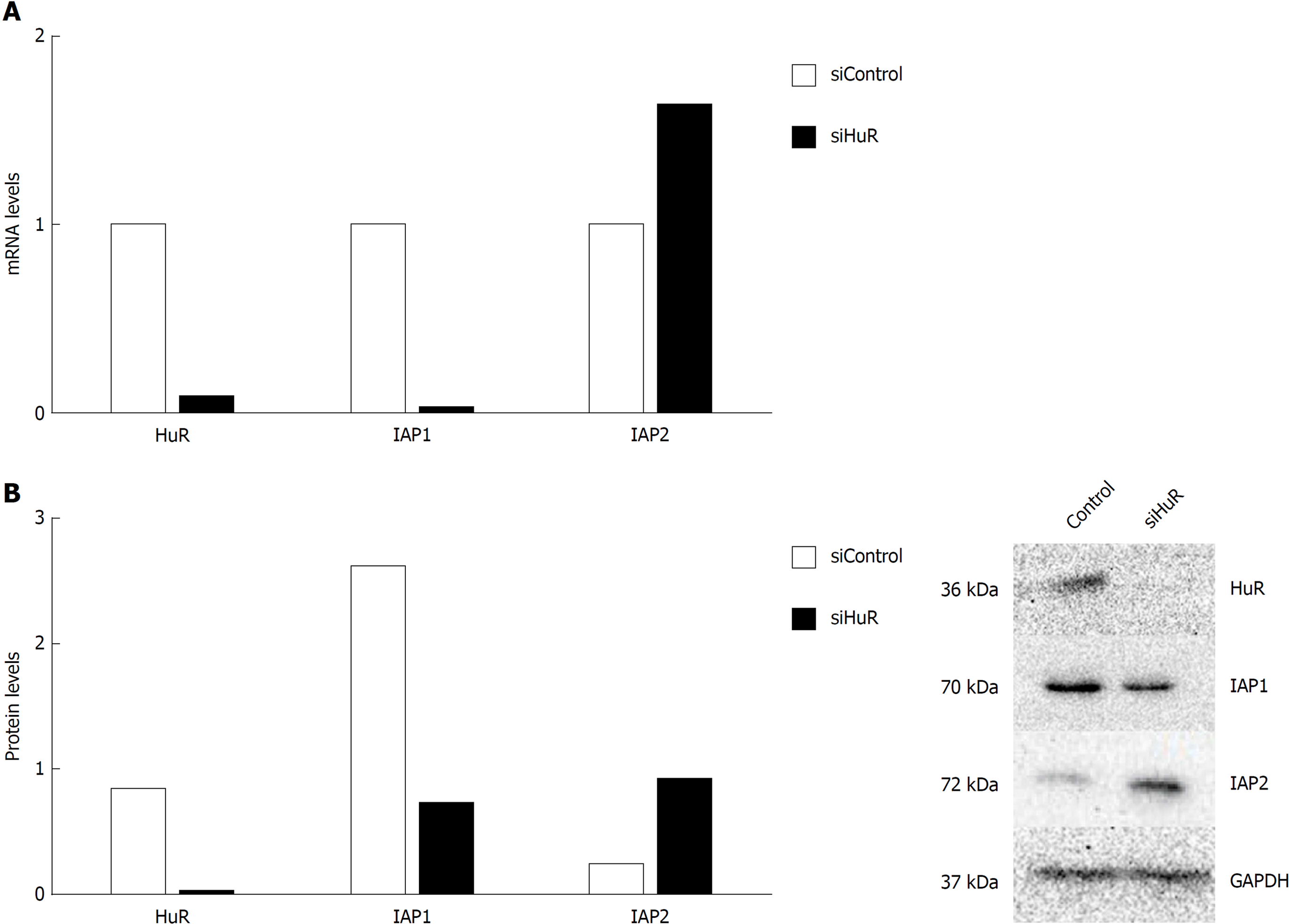
Figure 5 Human antigen R silencing is associated with inhibitors of apoptosis proteins mRNA and protein expression regulation in pancreatic cancer cells.
Human antigen R (HuR) silencing decreased inhibitor of apoptosis protein 1 (IAP1) and increased inhibitors of apoptosis protein 2 (IAP2) mRNA expression (A) and decreased IAP1 and increased IAP2 protein expression (B). IAP1: Inhibitor of apoptosis protein 1; IAP2: Inhibitor of apoptosis protein 2; HuR: Human antigen R; GAPDH: Glyceraldehyde-3-phosphate dehydrogenase.
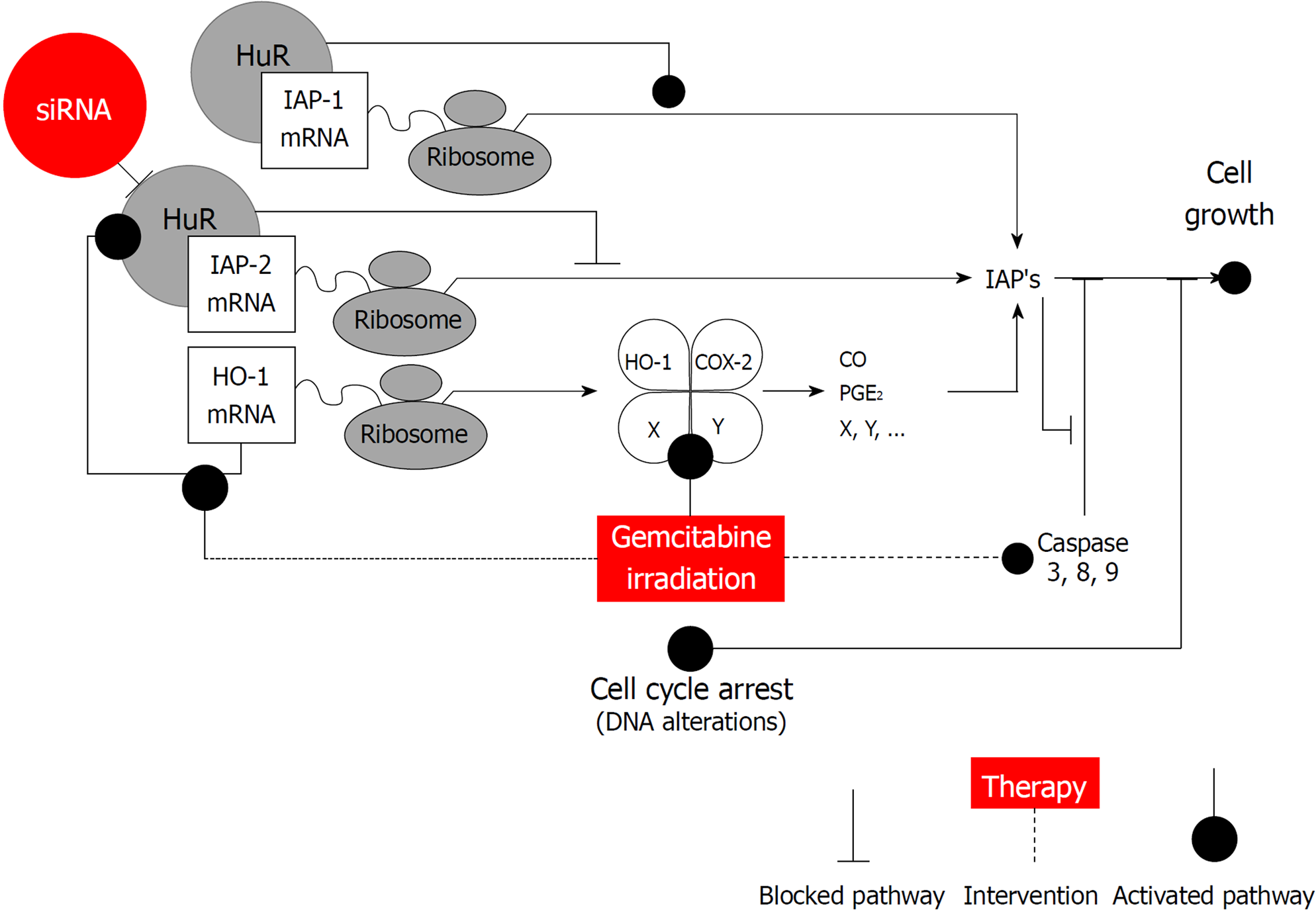
Figure 6 Human antigen R is an important regulator of pancreatic cancer cell growth and survival.
Human antigen R (HuR) can function through the regulation of the stability or translation of target mRNAs that encode multiple cancer-related proteins as inhibitors of apoptosis proteins (IAPs), heme oxygenase-1 (HO-1), cyclooxygenase-2 (COX-2). HuR can bind to IAPs and HO-1 and stabilize them, leading to an increased cell growth. However, when HuR is silenced, IAP2 overexpression is altered that could be modulated by HO-1 and a production of the COX-2 metabolites: prostaglandin E2 and carbon monoxide. Additionally, IAPs inhibit caspase activation and/or activity. IAPs: Inhibitors of apoptosis proteins; HuR: Human antigen R; HO-1: Heme oxygenase-1; COX-2: Cyclooxygenase-2; PGE2: Prostaglandin E2; CO: Carbon monoxide.
- Citation: Lukosiute-Urboniene A, Jasukaitiene A, Silkuniene G, Barauskas V, Gulbinas A, Dambrauskas Z. Human antigen R mediated post-transcriptional regulation of inhibitors of apoptosis proteins in pancreatic cancer. World J Gastroenterol 2019; 25(2): 205-219
- URL: https://www.wjgnet.com/1007-9327/full/v25/i2/205.htm
- DOI: https://dx.doi.org/10.3748/wjg.v25.i2.205


