Copyright
©The Author(s) 2019.
World J Gastroenterol. May 7, 2019; 25(17): 2045-2057
Published online May 7, 2019. doi: 10.3748/wjg.v25.i17.2045
Published online May 7, 2019. doi: 10.3748/wjg.v25.i17.2045
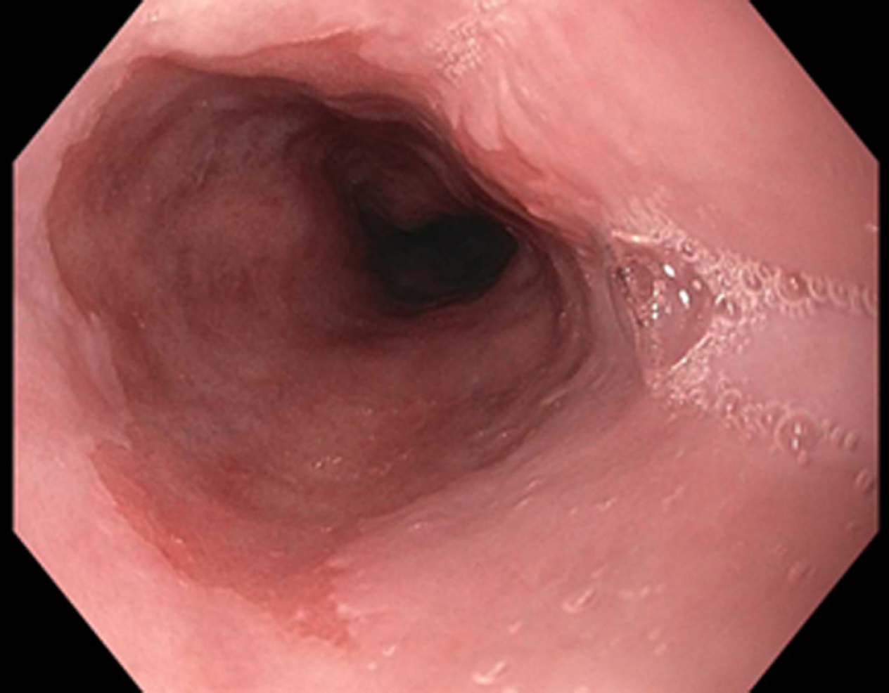
Figure 1 Barrett’s esophagus segment under while light high definition endoscopy.
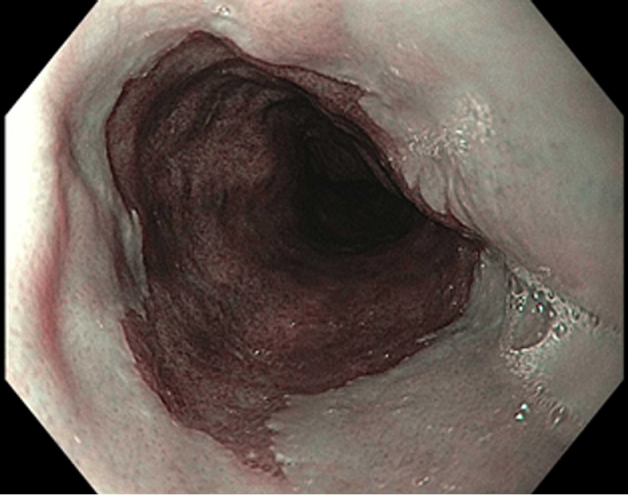
Figure 2 Barrett’s esophagus using narrow band imaging.
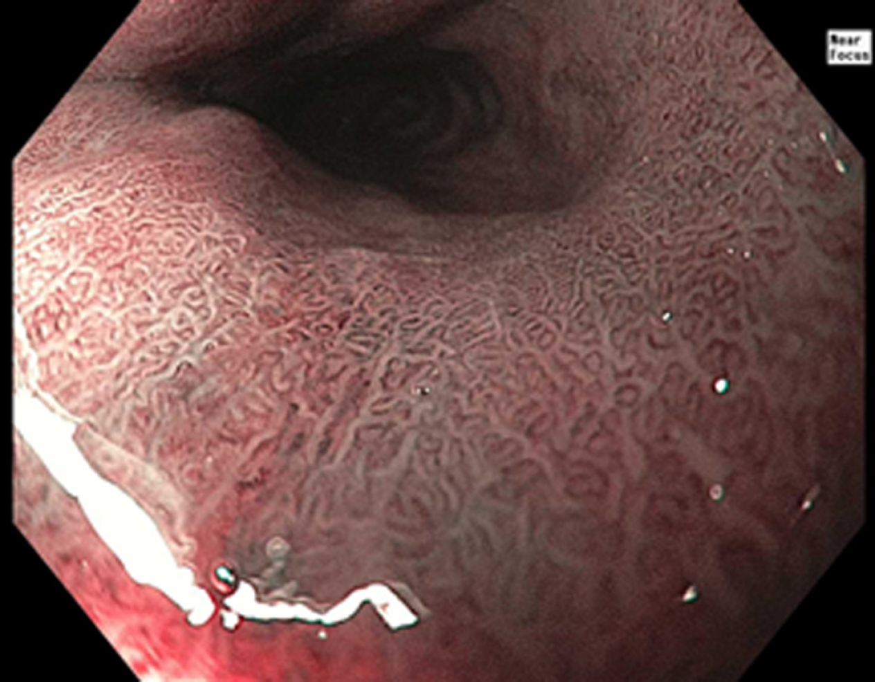
Figure 3 Barrett’s esophagus using zoom magnification endoscopy (near focus).
Figure 4 Confocal endomicroscopy imaging.
A: Barrett’s esophagus with intestinal metaplasia; B: Barrett’s esophagus with high grade dysplasia; C: Esophageal adenocarcinoma.
- Citation: Steele D, Baig KKK, Peter S. Evolving screening and surveillance techniques for Barrett's esophagus. World J Gastroenterol 2019; 25(17): 2045-2057
- URL: https://www.wjgnet.com/1007-9327/full/v25/i17/2045.htm
- DOI: https://dx.doi.org/10.3748/wjg.v25.i17.2045


