Copyright
©The Author(s) 2019.
World J Gastroenterol. Apr 21, 2019; 25(15): 1899-1906
Published online Apr 21, 2019. doi: 10.3748/wjg.v25.i15.1899
Published online Apr 21, 2019. doi: 10.3748/wjg.v25.i15.1899
Figure 1 Upper gastrointestinal endoscopy and tissue biopsy diagnosis.
A: Upper gastrointestinal endoscopy showed multiple gastric ulcers in the vestibular area. B: Biopsy tissue diagnosis showed large cells with intranuclear inclusions on hematoxylin and eosin staining (arrows) (× 200). C: Cytomegalovirus positive cells were observed through immunostaining (× 200).
Figure 2 Lower gastrointestinal endoscopy and tissue biopsy diagnosis.
A: Lower gastrointestinal showed a deep ulcer in the transverse colon. B: Biopsy tissue diagnosis showed large cells with intranuclear inclusions on hematoxylin and eosin staining (arrow) (× 400). C: Cytomegalovirus positive cells were observed through immunostaining (× 400).
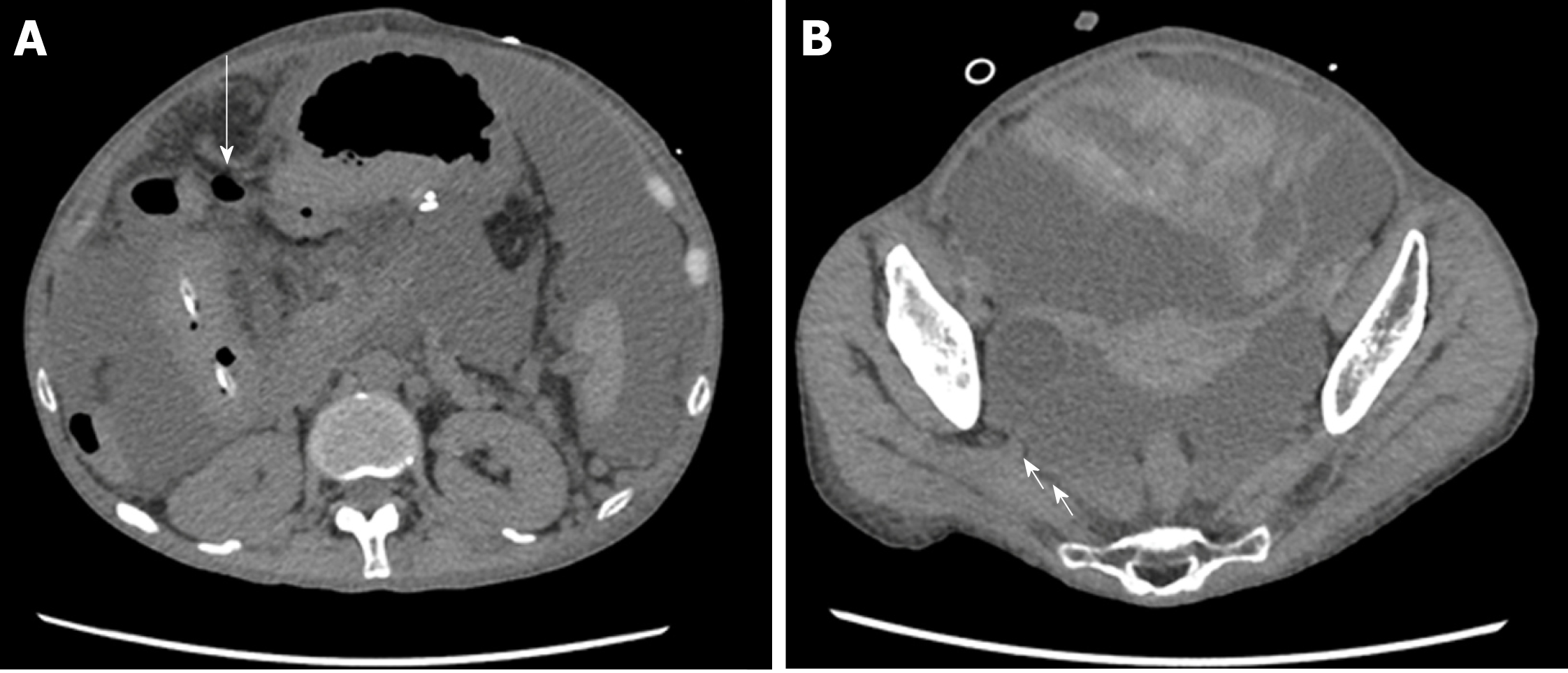
Figure 3 Abdominal computed tomography images.
A: Abdominal computed tomography (CT) revealed intraperitoneal free air near the transverse colon (arrow). B: Abdominal CT revealed hemorrhagic ascites in the pelvis (arrow).
- Citation: Yokose T, Obara H, Shinoda M, Nakano Y, Kitago M, Yagi H, Abe Y, Yamada Y, Matsubara K, Oshima G, Hori S, Ibuki S, Higashi H, Masuda Y, Hayashi M, Mori T, Kawaida M, Fujimura T, Hoshino K, Kameyama K, Kuroda T, Kitagawa Y. Colon perforation due to antigenemia-negative cytomegalovirus gastroenteritis after liver transplantation: A case report and review of literature. World J Gastroenterol 2019; 25(15): 1899-1906
- URL: https://www.wjgnet.com/1007-9327/full/v25/i15/1899.htm
- DOI: https://dx.doi.org/10.3748/wjg.v25.i15.1899


