Copyright
©The Author(s) 2018.
World J Gastroenterol. Feb 28, 2018; 24(8): 949-956
Published online Feb 28, 2018. doi: 10.3748/wjg.v24.i8.949
Published online Feb 28, 2018. doi: 10.3748/wjg.v24.i8.949
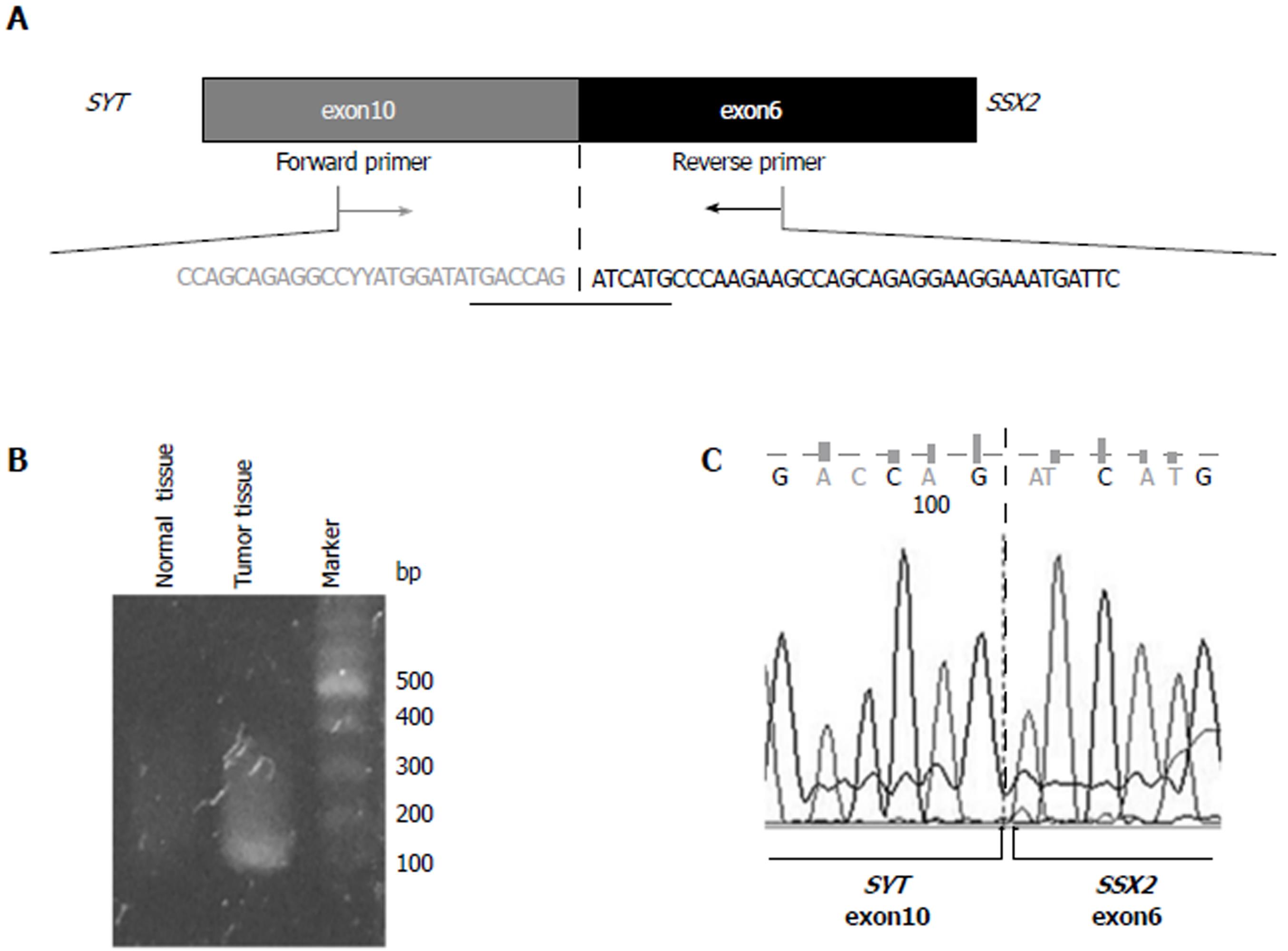
Figure 1 Identification of the SYT-SSX2 fusion transcript.
A: Structure of the SYT-SSX2 fusion transcript. Exon 10 of the SYT gene and exon 6 of the SSX2 gene are fused together in this transcript. The sequencing of exon is shown. Forward and reverse primers were designed in exon 10 of the SYT gene and exon 6 of the SSX2 gene, respectively; B: The RT-PCR product, including the SYT-SSX2 fusion site, was obtained from tumor tissue samples only. This analysis was performed more than three times, and a representative result of an electrophoresis is shown; C: The result of direct sequencing of the PCR product is shown. Exon 10 of the SYT gene is fused to exon 6 of the SSX2 gene.
Figure 2 Detection of the SYT-SSX2 fusion sequence.
A: The intronic primer settings in the SYT and SSX2 genes are shown; B: Genomic PCR products were obtained from tumor tissue samples only. This analysis was performed more than two times, and a representative result of an electrophoresis is shown; C: The intronic breakpoint was confirmed by direct sequencing.
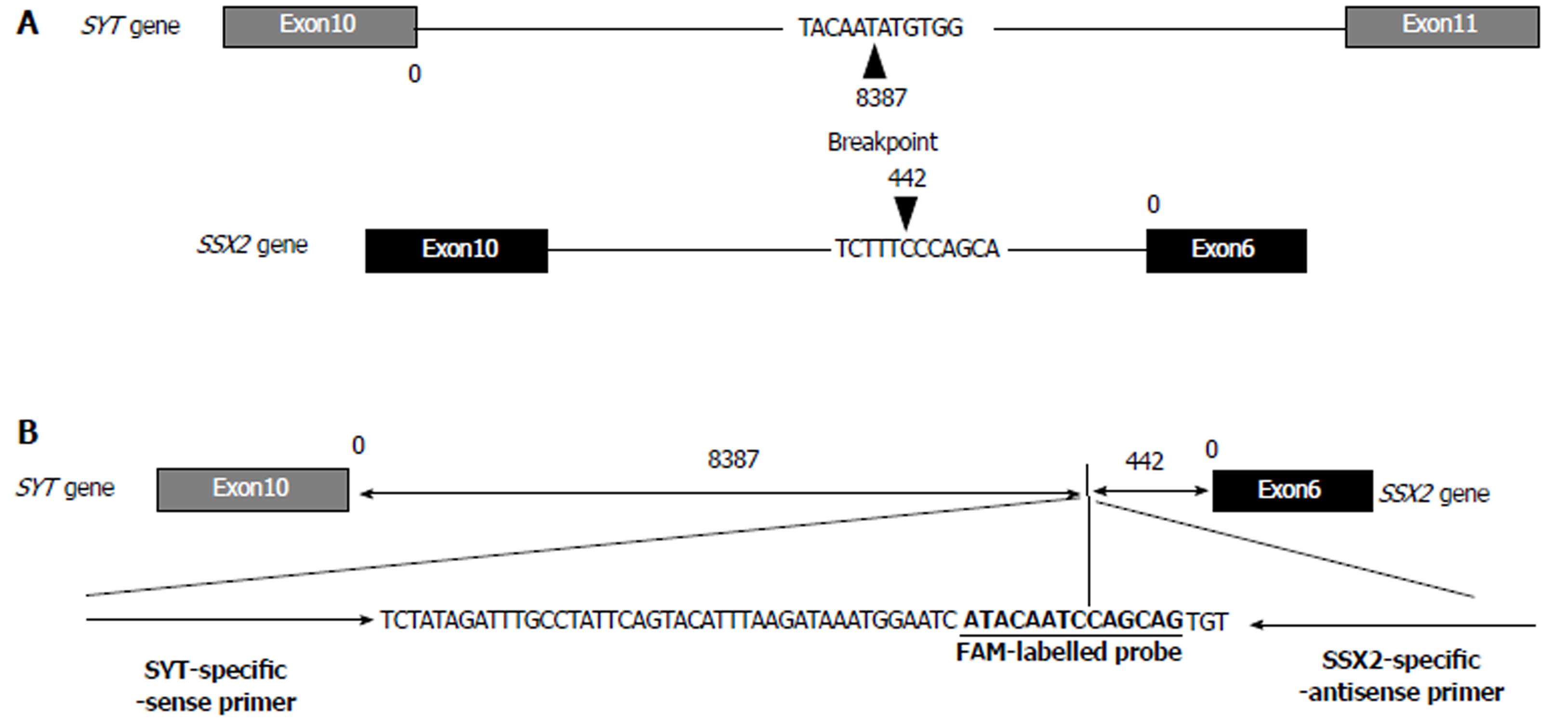
Figure 3 Intronic breakpoint and structure of the fusion sequence with specific probe and primer sets.
A: The arrowheads indicate the position and sequence of the intronic breakpoint of the SYT and SSX2 genes; B: The structure and sequence of the FAM-labeled probe and primer sets specific to the intronic breakpoint of the SYT-SSX fusion sequence.
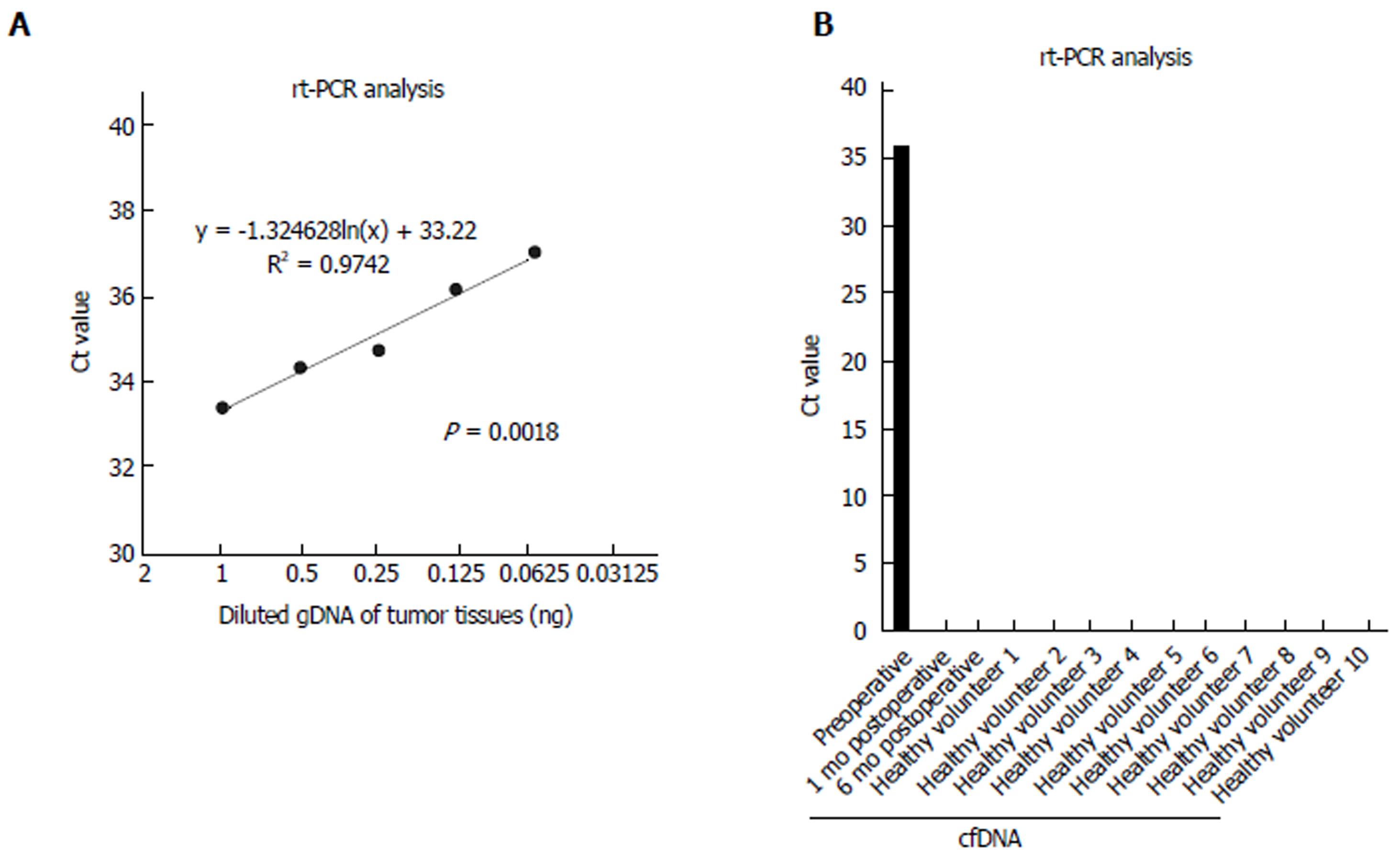
Figure 4 Detection of the fusion gene sequence in cell-free DNA using specific probe and primer sets.
A: The range of reproducibility of rt-PCR with the specific probe and primer sets was confirmed. Diluted serial tumor gDNA of 1-0.063 ng was used. The dotted line indicates an approximate curve (R² = 0.9742, P = 0.0018); B: cfDNA samples of a patient and 10 healthy volunteers were analyzed using rt-PCR. rt-PCR was performed in duplicate, and mean Ct values are indicated. Standard deviation was calculated from duplicate samples. cfDNA: Cell-free DNA.
- Citation: Ogino S, Konishi H, Ichikawa D, Hamada J, Shoda K, Arita T, Komatsu S, Shiozaki A, Okamoto K, Yamazaki S, Yasukawa S, Konishi E, Otsuji E. Detection of fusion gene in cell-free DNA of a gastric synovial sarcoma. World J Gastroenterol 2018; 24(8): 949-956
- URL: https://www.wjgnet.com/1007-9327/full/v24/i8/949.htm
- DOI: https://dx.doi.org/10.3748/wjg.v24.i8.949


