Copyright
©The Author(s) 2018.
World J Gastroenterol. Feb 7, 2018; 24(5): 593-601
Published online Feb 7, 2018. doi: 10.3748/wjg.v24.i5.593
Published online Feb 7, 2018. doi: 10.3748/wjg.v24.i5.593
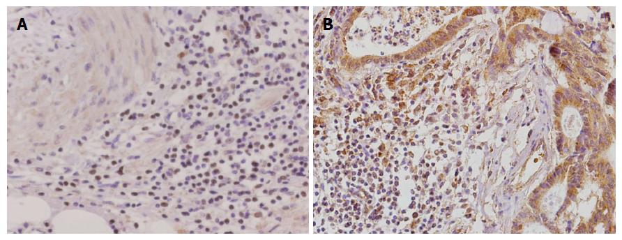
Figure 1 CD68 and Sema4D expression in gastric carcinoma (streptavidin-peroxidase, × 400).
A: Positive staining for CD68 in gastric carcinoma tissues; B: Positive staining for Sema4D in gastric carcinoma tissues.
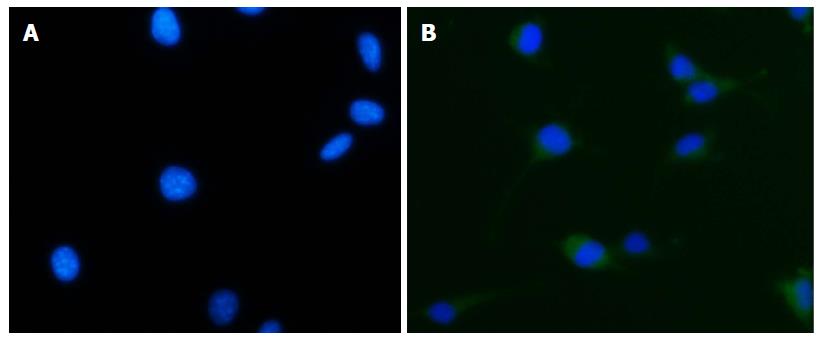
Figure 2 Immunofluorescence staining of cells (400 ×).
A: CD68-negative cells; B: CD68-positive cells.
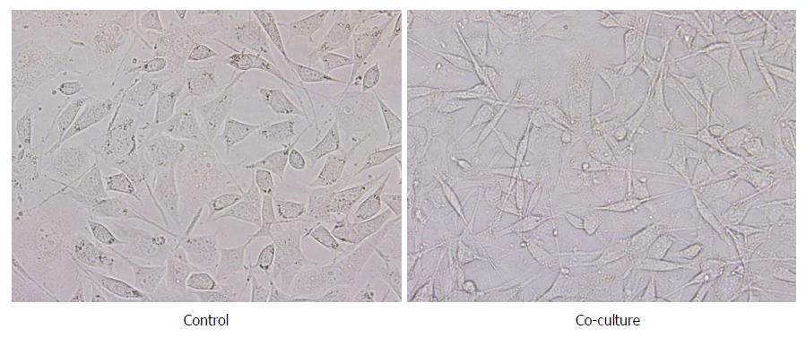
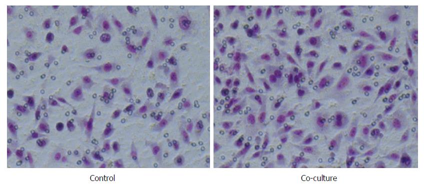
Figure 3 Morphological changes of gastric carcinoma SGC-7901 cells.
A: Control; B: Co-culture group (400 ×).
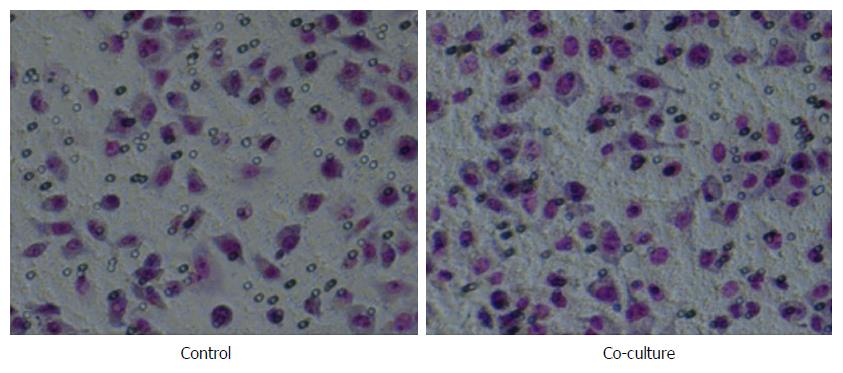
Figure 4 Tumor-associated macrophages promote SGC-7901 cell migration (200 ×, P < 0.
01).
Figure 5 Tumor-associated macrophages promote SGC-7901 cell invasion (200 ×, P < 0.
01).
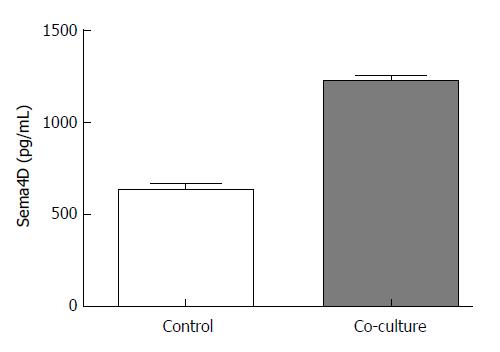
Figure 6 Expression of Sema4D protein in SGC-7901 cell supernatants of the control and co-culture experimental groups.
- Citation: Li H, Wang JS, Mu LJ, Shan KS, Li LP, Zhou YB. Promotion of Sema4D expression by tumor-associated macrophages: Significance in gastric carcinoma. World J Gastroenterol 2018; 24(5): 593-601
- URL: https://www.wjgnet.com/1007-9327/full/v24/i5/593.htm
- DOI: https://dx.doi.org/10.3748/wjg.v24.i5.593


