Copyright
©The Author(s) 2018.
World J Gastroenterol. Dec 21, 2018; 24(47): 5379-5390
Published online Dec 21, 2018. doi: 10.3748/wjg.v24.i47.5379
Published online Dec 21, 2018. doi: 10.3748/wjg.v24.i47.5379
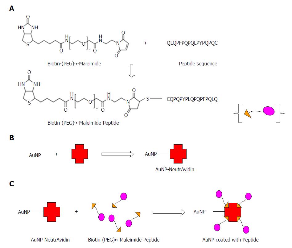
Figure 1 Schematic representation of preparation of peptide coated AuNPs.
A: Maleimide groups of the linker reacted specifically with free (reduced) sulfhydryl’s in the peptide sequence to form stable thio-ether bonds; B: NeutrAvidin was coated on the surface of the AuNPs to obtain NeutrAvidin-AuNP particles; C: The biotin end of the linker interacted with the NeutrAvidin-AuNPs resulting in the formation of peptide coated AuNPs.
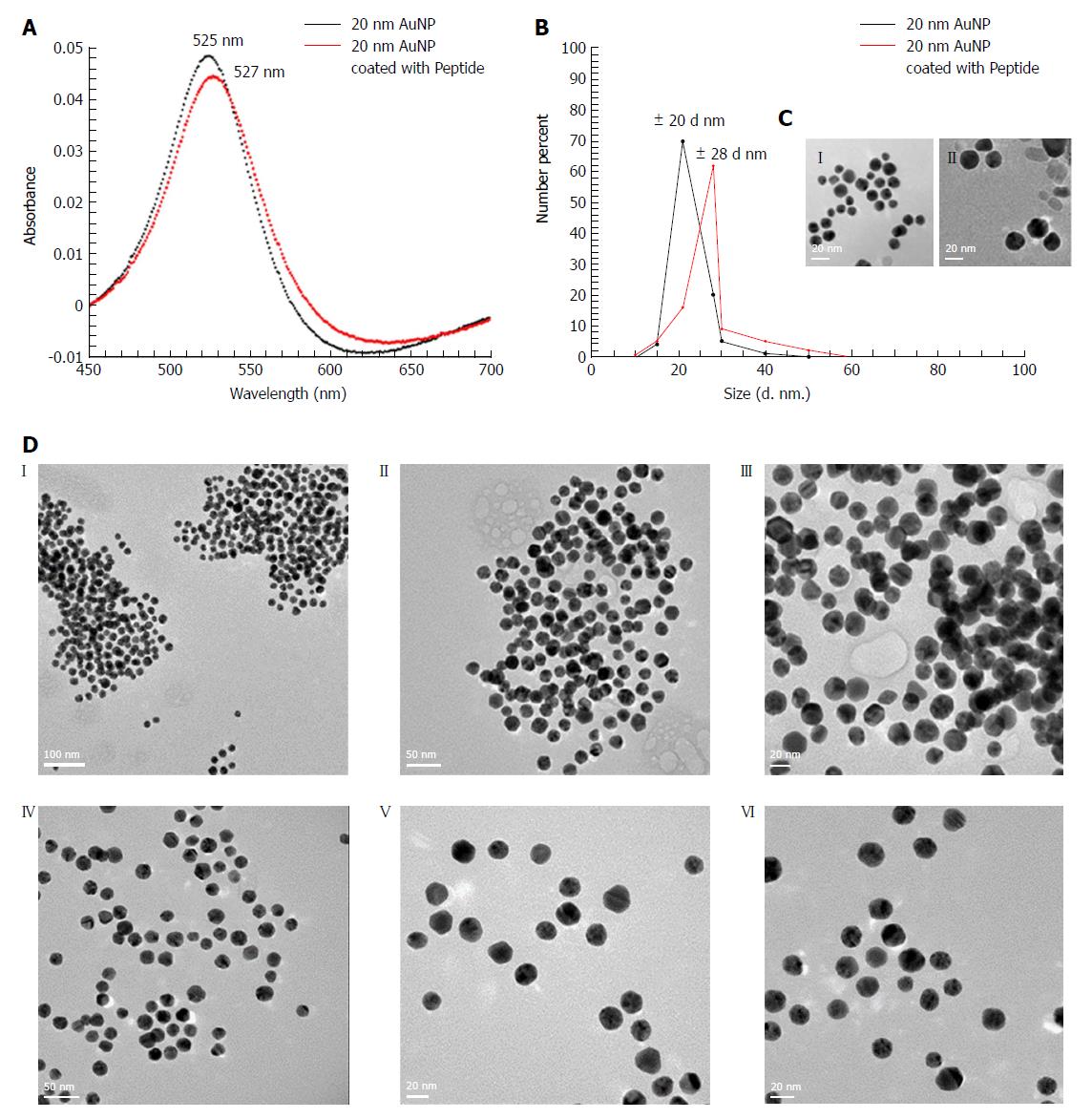
Figure 2 Characterization of peptide coated AuNPs.
A: Characterization of AuNP coated with peptide using a UV-Vis spectrophotometer indicating a spectral red shift in wavelength from 525 nm (for 20 nm AuNP only) to 527 nm (for 20 nm AuNP coated with peptide); B: Characterization of AuNP coated with peptide using DLS that showed an increase in the hydrodynamic size of the uncoated vs coated particles from 20 nm to 28 nm respectively; C: High resolution TEM images of (1) uncoated AuNPs, (2) AuNPs coated with peptide showing a “halo” layer surrounding the surface of the nanoparticles indicating coating of the gold with the peptide had occurred. In contrast, the “halo” effect was not observed on the surface of the un-coated AuNPs; D: High resolution TEM images following the incubation with AGA (12 μg/mL) (1, 2, 3) AuNPs coated with peptide showing aggregation confirming coating of peptide on AuNP (4, 5, 6) Uncoated AuNPs remained dispersed. DLS: Dynamic light scattering; TEM: Transmission electron microscopy.
Figure 3 Testing peptide-coated AuNPs with anti-gliadin.
A: Reduction in color from red to translucent and absorbance was observed in AuNP coated with peptide (S) and incubated with AGA at various dilutions (S1) 2 μg/mL, (S2) 4 μg/mL, (S3) 6 μg/mL, (S4) 8 μg/mL, (S5) 10 μg/mL), (S6) 12 μg/mL, (S7) 14 μg/mL, (S8) 16 μg/mL, (S9) 18 µg/mL and (S10) 20 μg/mL. No significant reduction in color or shift in peak wavelength was observed in peptide coated AuNP incubated with control rabbit IgG at dilutions (C1) 2 μg/mL, (C2) 4 μg/mL, (C3) 6 μg/mL, (C4) 8 μg/mL, (C5) 10 μg/mL), (C6) 12 μg/mL, (C7) 14 μg/mL, (C8) 16 μg/mL, (C9) 18 μg/mL and (C10) 20 μg/mL; B: Representation of specificity based on UV-Vis absorbance spectra for the antibody interactions at equal concentrations of AGA and control antibody; C: Colorimetric response curve plotted on AuNP coated with peptide following the addition of AGA at different dilutions. AGA: Anti-gliadin.
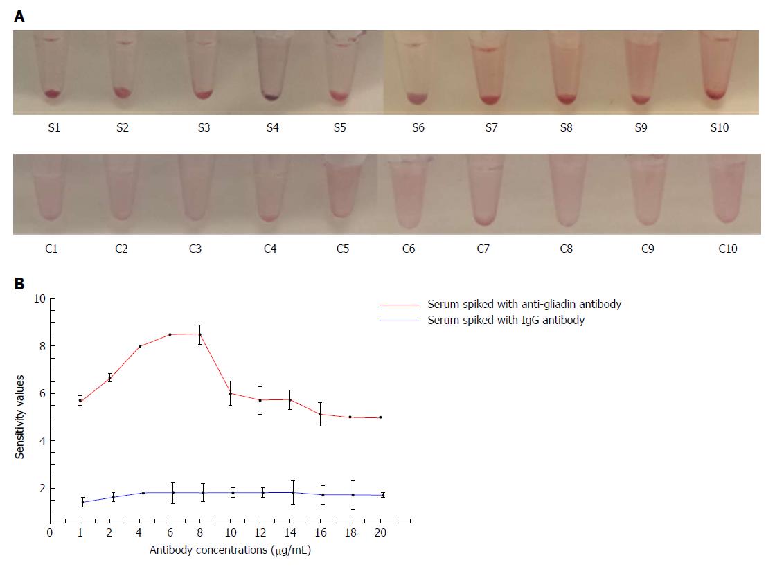
Figure 4 Detection of anti-gliadin in spiked human serum using peptide-coated AuNPs.
A: Reduction in color from red to translucent as well as precipitate formation was observed in AuNP coated with peptide in the presence of AGA and serum at different concentrations (S1-S10). No reduction in color from red to translucent or precipitate formation was observed in AuNP coated with peptide in the presence of control IgG and serum (C1-C10); B: Colorimetric response curve plotted in AuNP coated with peptide in 1:20 diluted serum following the addition of AGA at different dilutions. AGA: Anti-gliadin.
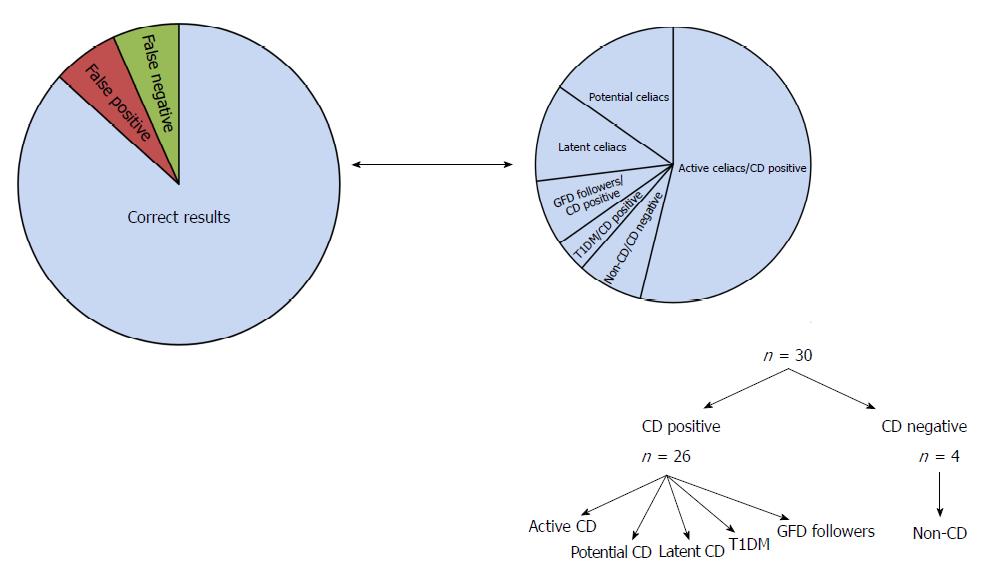
Figure 5 Representation of the distribution of clinical samples using AuNP-peptide-anti-gliadin test.
- Citation: Kaur A, Shimoni O, Wallach M. Novel screening test for celiac disease using peptide functionalised gold nanoparticles. World J Gastroenterol 2018; 24(47): 5379-5390
- URL: https://www.wjgnet.com/1007-9327/full/v24/i47/5379.htm
- DOI: https://dx.doi.org/10.3748/wjg.v24.i47.5379


