Copyright
©The Author(s) 2018.
World J Gastroenterol. Nov 14, 2018; 24(42): 4728-4737
Published online Nov 14, 2018. doi: 10.3748/wjg.v24.i42.4728
Published online Nov 14, 2018. doi: 10.3748/wjg.v24.i42.4728
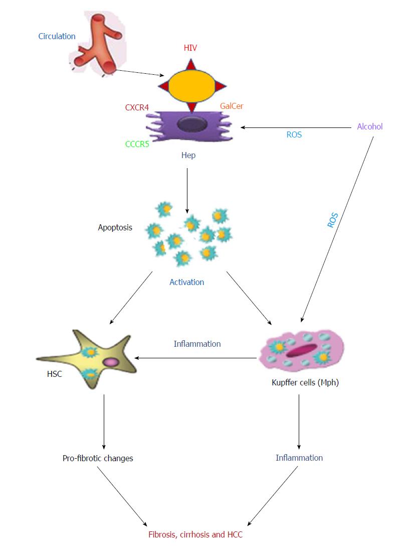
Figure 1 Possible mechanisms of interaction between human immunodeficiency virus-infected liver parenchymal and non-parenchymal cells.
Human immunodeficiency virus (HIV) infected immune cells are trapped by the liver. HIV envelope proteins interact with hepatocytes using the co-receptors CXCR4/CCR5 or GalCer to induce apoptosis. Apoptotic bodies from infected hepatocytes are captured by both hepatic stellate cells (HSC) and Kupffer cells. This process activates both cell types, which induce the profibrotic changes and inflammation, respectively. In addition, activated Kupffer cells, in turn, regulate HSCs activation. The second hit, alcohol, potentiates inflammation and fibrosis development by oxidative stress-induced hepatocyte apoptosis enhanced by HIV-infected Kupffer cells. All these combined events may lead to fibrosis, cirrhosis and hepatocellular carcinoma development. All HIV related events can be suppressed by antiretroviral therapy. HIV: Human immunodeficiency virus; ART: Antiretroviral therapy; GalCer: Galactosyl ceramide; Hep: Hepatocytes; ROS: Reactive oxygen species; HCC: Hepatocellular carcinoma.
- Citation: Ganesan M, Poluektova LY, Kharbanda KK, Osna NA. Liver as a target of human immunodeficiency virus infection. World J Gastroenterol 2018; 24(42): 4728-4737
- URL: https://www.wjgnet.com/1007-9327/full/v24/i42/4728.htm
- DOI: https://dx.doi.org/10.3748/wjg.v24.i42.4728


