Copyright
©The Author(s) 2018.
World J Gastroenterol. Nov 7, 2018; 24(41): 4698-4707
Published online Nov 7, 2018. doi: 10.3748/wjg.v24.i41.4698
Published online Nov 7, 2018. doi: 10.3748/wjg.v24.i41.4698
Figure 1 Superficially invasive T1b oesophageal adenocarcinoma.
A: Low power overview of the lesion. A moderately differentiated oesophageal adenocarcinoma is located at the squamocolumnar junction. This is a piecemeal excision of this lesion. The deep margin is clear (R0). There was no lymphovascular invasion or poor differentiation. Boxed area shown in B. B: High power of the invasive front showing a tumour gland just penetrating the original muscularis mucosae (asterisk).
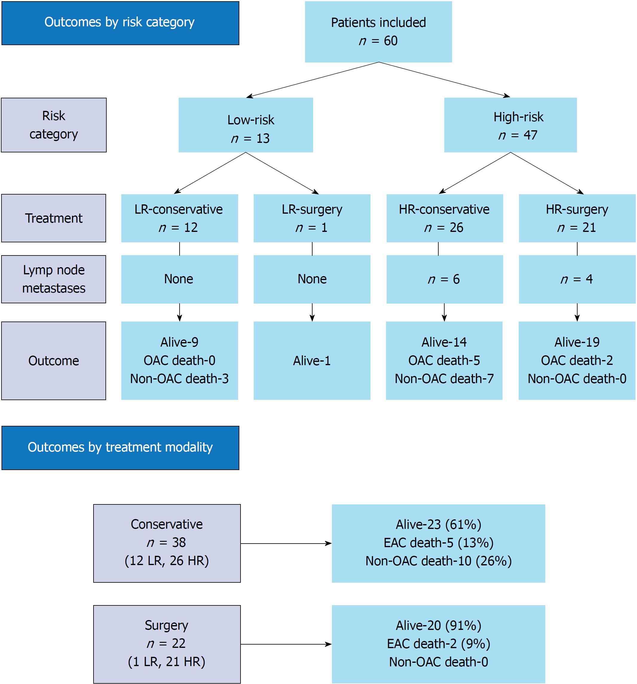
Figure 2 Flowchart depicting patient subgroups and outcomes.
Overall median follow-up 41 mo (IQR 26-53). OAC: Oesophageal adenocarcinoma; LR: Low-risk tumour; HR: High-risk tumour; IQR: Interquartile range.
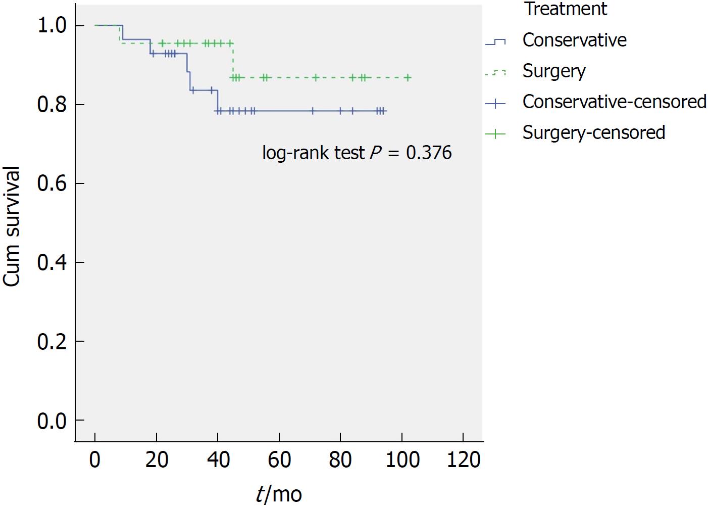
Figure 3 Kaplan-Meier disease-specific survival curve of all patients treated conservatively and surgically.
There was no statistically significant difference (excluded were 10 patients from conservative group who died of non-oesophageal adenocarcinoma-related cause).
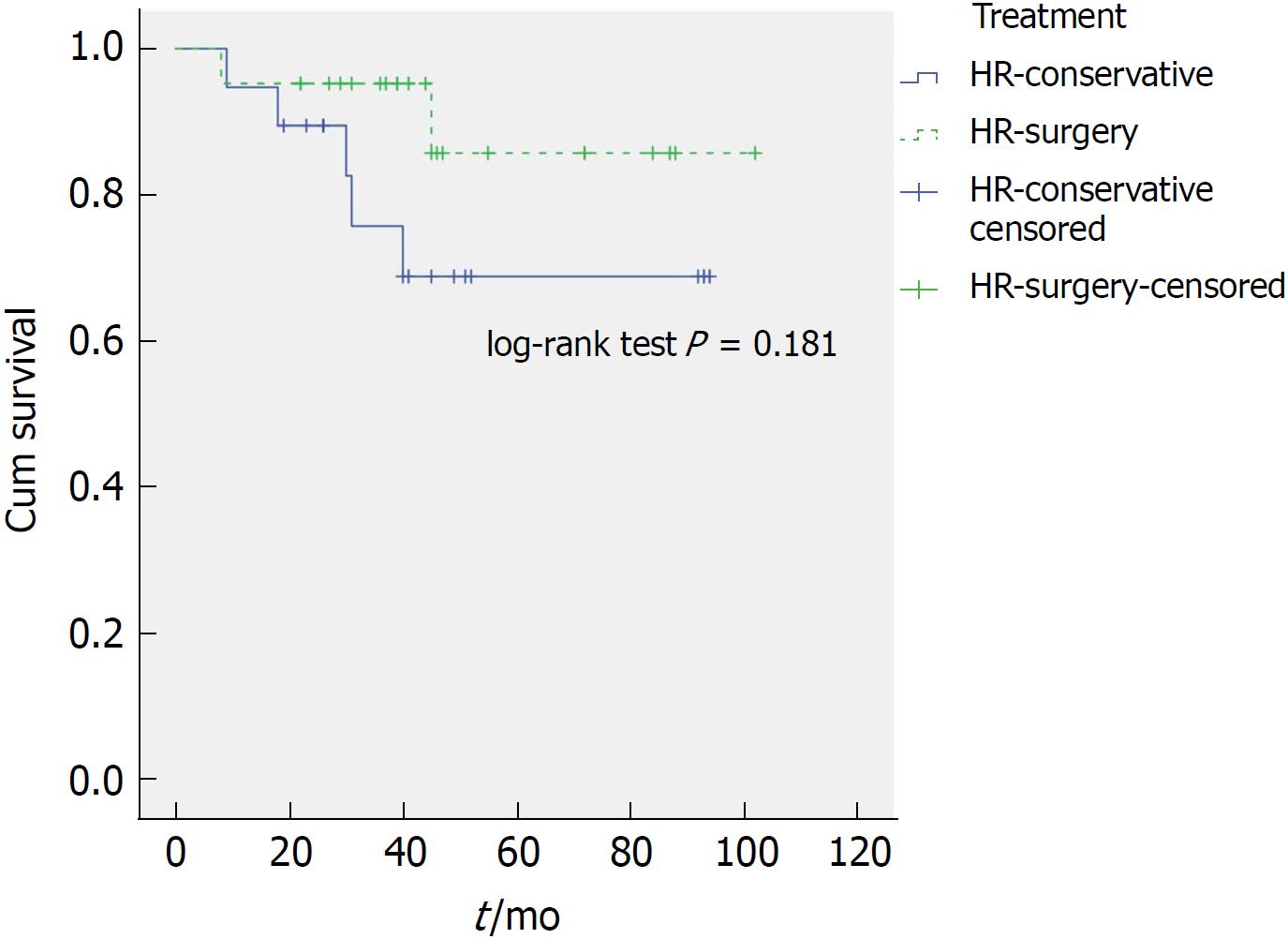
Figure 4 Kaplan-Meier disease-specific survival curve of patients with high-risk tumours treated conservatively and surgically (excluded were 7 patients from high-risk-conservative group who died of non-oesophageal adenocarcinoma-related cause).
There was no statistically significant difference, although a trend towards a better survival of surgery patients is observed.
- Citation: Graham D, Sever N, Magee C, Waddingham W, Banks M, Sweis R, Al-Yousuf H, Mitchison M, Alzoubaidi D, Rodriguez-Justo M, Lovat L, Novelli M, Jansen M, Haidry R. Risk of lymph node metastases in patients with T1b oesophageal adenocarcinoma: A retrospective single centre experience. World J Gastroenterol 2018; 24(41): 4698-4707
- URL: https://www.wjgnet.com/1007-9327/full/v24/i41/4698.htm
- DOI: https://dx.doi.org/10.3748/wjg.v24.i41.4698


