Copyright
©The Author(s) 2018.
World J Gastroenterol. Oct 14, 2018; 24(38): 4412-4418
Published online Oct 14, 2018. doi: 10.3748/wjg.v24.i38.4412
Published online Oct 14, 2018. doi: 10.3748/wjg.v24.i38.4412
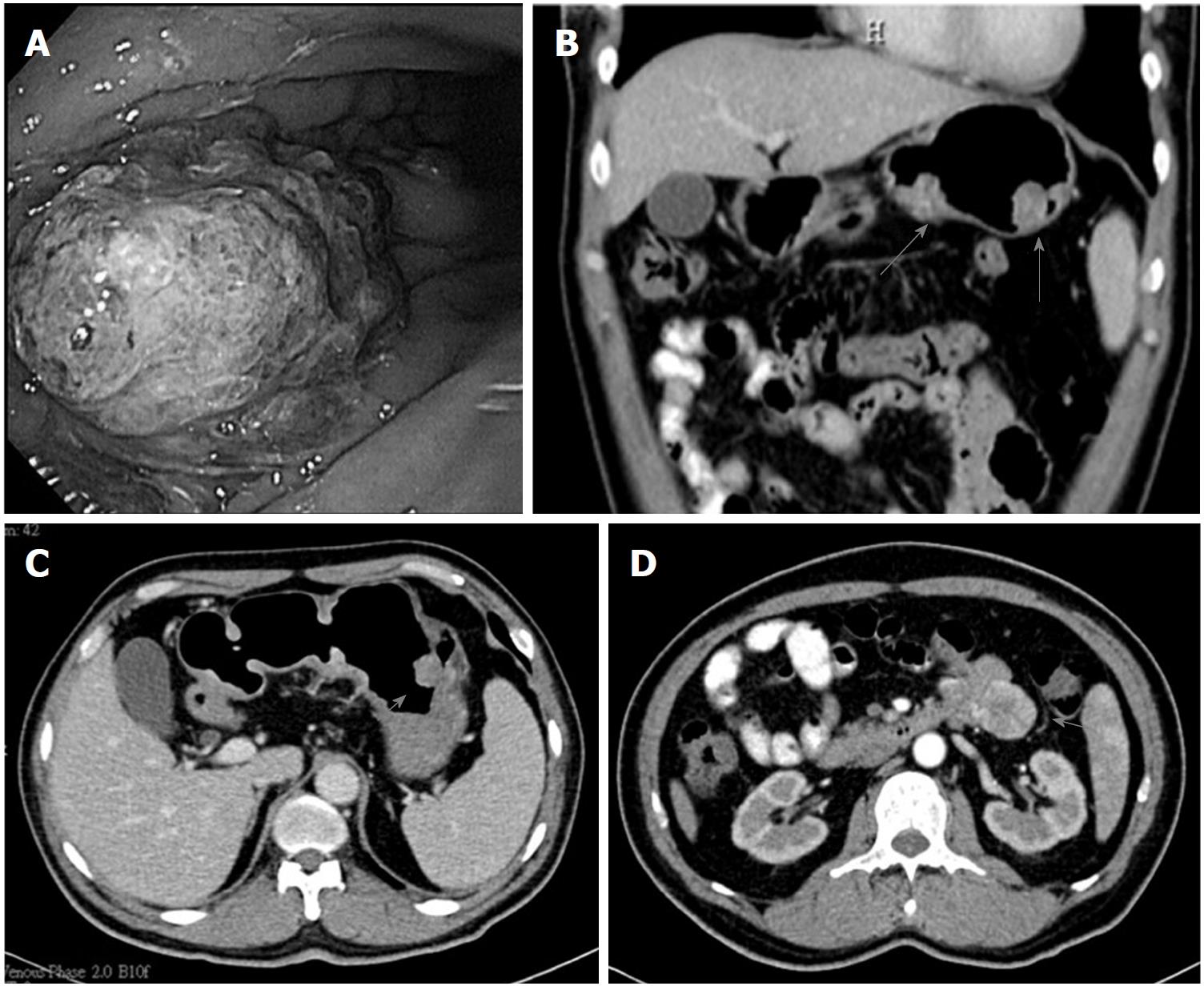
Figure 1 Endoscopic findings and computed tomography scans of the abdomen of the patient at the diagnosis of gastric cancer.
A: Multiple polyps in the gastric body and duodenal bulb were demonstrated by EGD; B: The coronal and C: sagittal views from the CT scan; D: An exophytic mass measuring about 3.7 cm × 3.5 cm was incidentally found in the proximal jejunum by CT. CT: Computed tomography; EGD: Esophagogastroduodenoscopy.
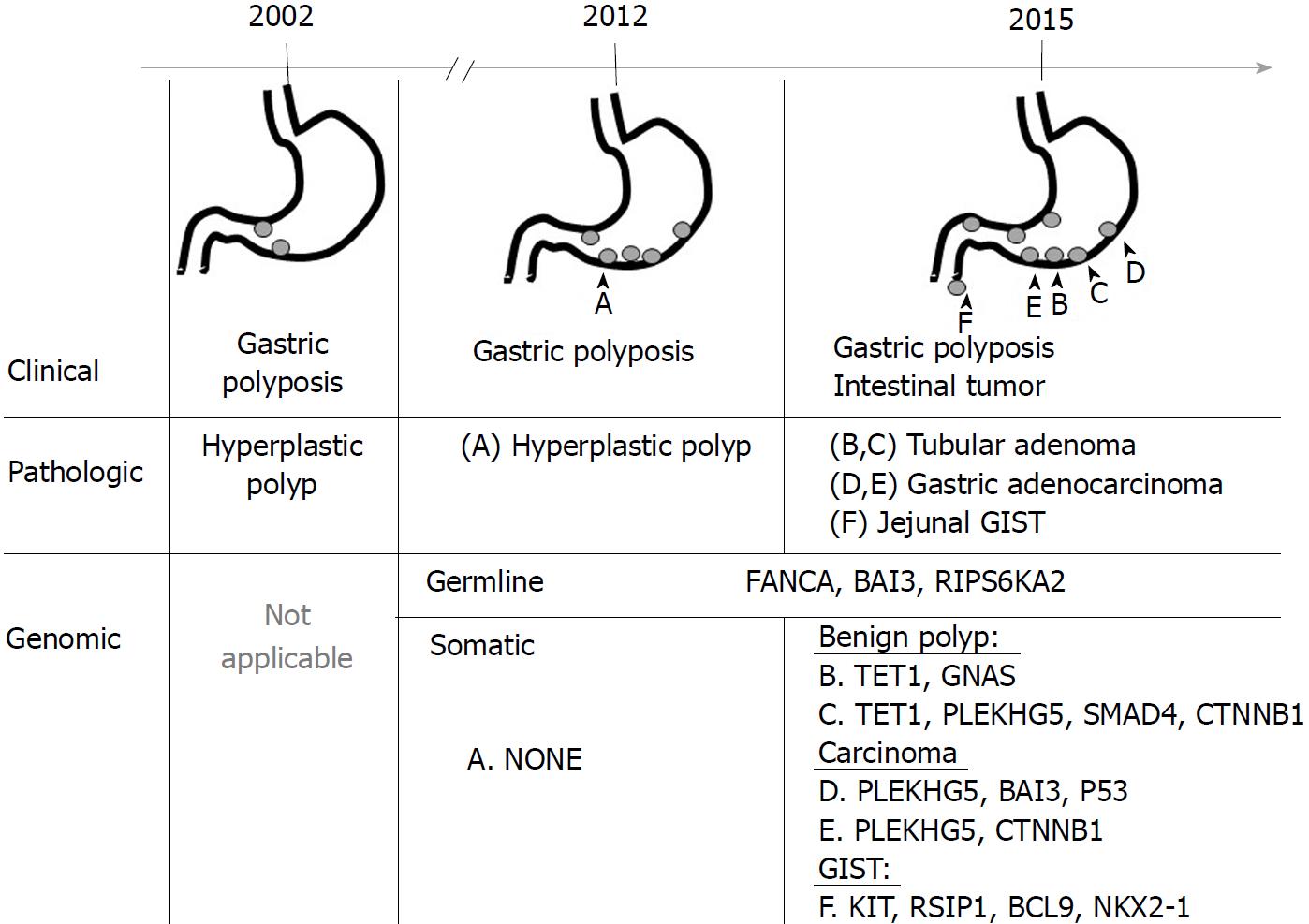
Figure 2 Chronological summary of the clinical, pathologic, and genomic findings of gastric polyps and tumor specimen labeling (A-F) for massive parallel sequencing.
GIST: Gastro-intestinal stromal tumor.
- Citation: Huang JP, Lin J, Tzen CY, Huang WY, Tsai CC, Chen CJ, Lu YJ, Chou KF, Su YW. FANCA D1359Y mutation in a patient with gastric polyposis and cancer susceptibility: A case report and review of literature. World J Gastroenterol 2018; 24(38): 4412-4418
- URL: https://www.wjgnet.com/1007-9327/full/v24/i38/4412.htm
- DOI: https://dx.doi.org/10.3748/wjg.v24.i38.4412


