Copyright
©The Author(s) 2018.
World J Gastroenterol. Sep 21, 2018; 24(35): 4077-4085
Published online Sep 21, 2018. doi: 10.3748/wjg.v24.i35.4077
Published online Sep 21, 2018. doi: 10.3748/wjg.v24.i35.4077
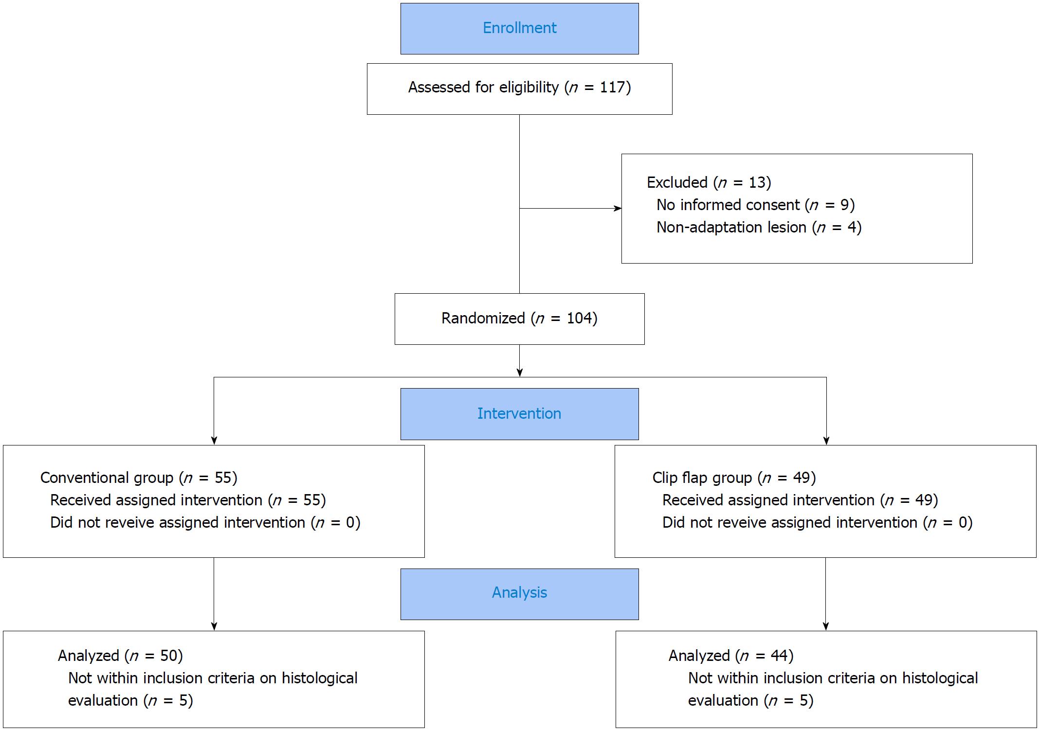
Figure 1 Flow diagram of this study.
We enrolled 117 patients who were scheduled to undergo ESD for gastric tumors from May 2015 to October 2016. A total of 104 patients were randomized to the conventional and the clip-flap groups. After ESD, ten patients had a lesion outside the inclusion criteria of early-stage gastric cancer. ESD: Endoscopic submucosal dissection.
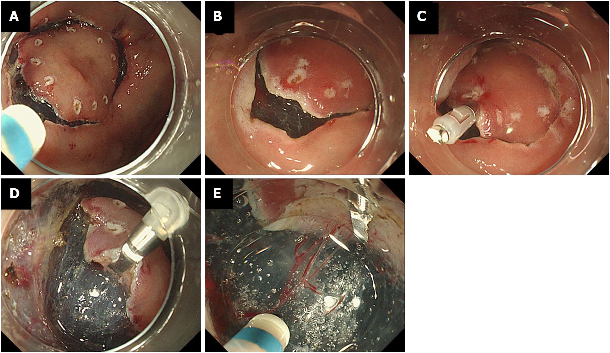
Figure 2 Clip-flap methods.
A: The mucosal circumference incision of gastric tumor is performed in the conventional manner; B: A deeper cut is made at the point attached the endoclip; C: The endoclip is attached to the exfoliated mucosa. The head of the endoclip falls slightly toward the gastric lumen, allowing the attachment to be easily inserted under the endoclip; D: The attachment is inserted under the endoclip, and then the mucosa and submucosal layer are elevated by the endoclip; E: The gastric tumor is dissected with the endoknife under direct vision.
- Citation: Ban H, Sugimoto M, Otsuka T, Murata M, Nakata T, Hasegawa H, Inatomi O, Bamba S, Andoh A. Usefulness of the clip-flap method of endoscopic submucosal dissection: A randomized controlled trial. World J Gastroenterol 2018; 24(35): 4077-4085
- URL: https://www.wjgnet.com/1007-9327/full/v24/i35/4077.htm
- DOI: https://dx.doi.org/10.3748/wjg.v24.i35.4077


