Copyright
©The Author(s) 2018.
World J Gastroenterol. Aug 28, 2018; 24(32): 3671-3676
Published online Aug 28, 2018. doi: 10.3748/wjg.v24.i32.3671
Published online Aug 28, 2018. doi: 10.3748/wjg.v24.i32.3671
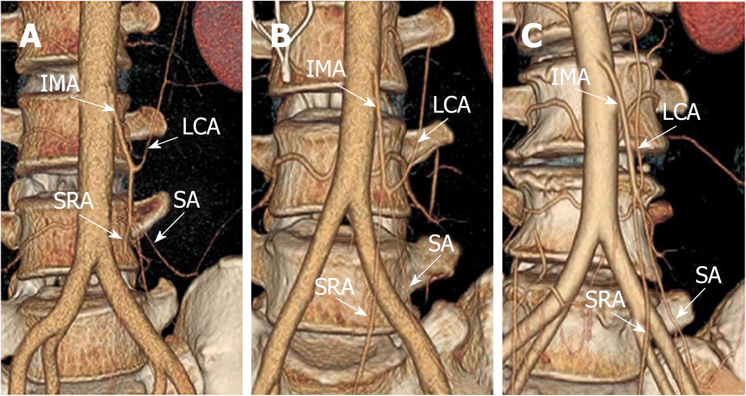
Figure 1 Preoperative 3D reconstruction of inferior mesenteric artery, left colic artery, sigmoid artery and superior rectal artery.
A: Type A, LCA arose independently from IMA. B: type B, LCA and SA branched from a common trunk from IMA. C: type C, LCA, SA, and SRA branched off at the same point. IMA: Inferior mesenteric artery; LCA: Left colic artery; SA: Sigmoid artery; SRA: Superior rectal artery.
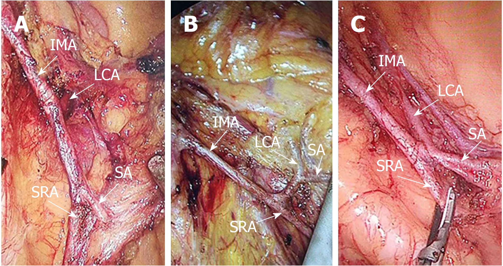
Figure 2 Inferior mesenteric artery, left colic artery, sigmoid artery and superior rectal artery in laparoscopic operation.
A: Type A, LCA arose independently from IMA. B: type B, LCA and SA branched from a common trunk from IMA. C: type C, LCA, SA, and SRA branched off at the same point. IMA: Inferior mesenteric artery; LCA: Left colic artery; SA: Sigmoid artery; SRA: Superior rectal artery.
Figure 3 Vascular schematic diagram of inferior mesenteric artery, left colic artery, sigmoid artery and superior rectal artery.
A: Type A, LCA arose independently from IMA. B: type B, LCA and SA branched from a common trunk from IMA. C: type C, LCA, SA, and SRA branched off at the same point. AA: Abdominal aorta; IMA: Inferior mesenteric artery; LCA: Left colic artery; SA: Sigmoid artery; SRA: Superior rectal artery.
- Citation: Wang KX, Cheng ZQ, Liu Z, Wang XY, Bi DS. Vascular anatomy of inferior mesenteric artery in laparoscopic radical resection with the preservation of left colic artery for rectal cancer. World J Gastroenterol 2018; 24(32): 3671-3676
- URL: https://www.wjgnet.com/1007-9327/full/v24/i32/3671.htm
- DOI: https://dx.doi.org/10.3748/wjg.v24.i32.3671


