Copyright
©The Author(s) 2018.
World J Gastroenterol. Jun 21, 2018; 24(23): 2501-2507
Published online Jun 21, 2018. doi: 10.3748/wjg.v24.i23.2501
Published online Jun 21, 2018. doi: 10.3748/wjg.v24.i23.2501
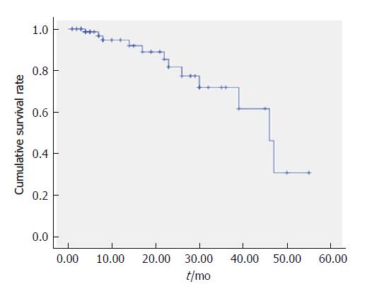
Figure 1 Kaplan-Meier curves of the overall survival for all 86 patients (months).
Figure 2 Images of case 1.
A: Angiography via the proper hepatic artery revealed a tumor stain in the liver; B: Portal trunk tumor thrombus; C: Super-selective catheterization and administered embolization and chemotherapy infusion; D: Post-operative computed tomography (CT) scan revealed scattered deposition of lipiodol inside the major portal vein tumor thrombosis as well as intra-hepatic lesions.
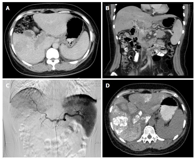
Figure 3 Images of case 2.
A: Pre-operative CT scan (right branch); B: Pre-operative CT scan (portal trunk); C: Tumor stain in the liver and right branch and a portal trunk tumor thrombus; D: Post-operative CT scan revealed the deposition of lipiodol inside the major portal vein tumor thrombosis.
- Citation: Zhu LZ, Xu S, Qian HL. Transarterial embolization and low-dose continuous hepatic arterial infusion chemotherapy with oxaliplatin and raltitrexed for hepatocellular carcinoma with major portal vein tumor thrombus. World J Gastroenterol 2018; 24(23): 2501-2507
- URL: https://www.wjgnet.com/1007-9327/full/v24/i23/2501.htm
- DOI: https://dx.doi.org/10.3748/wjg.v24.i23.2501


