Copyright
©The Author(s) 2018.
World J Gastroenterol. May 7, 2018; 24(17): 1911-1918
Published online May 7, 2018. doi: 10.3748/wjg.v24.i17.1911
Published online May 7, 2018. doi: 10.3748/wjg.v24.i17.1911
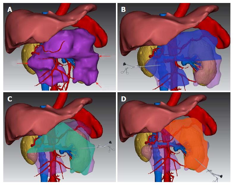
Figure 1 Three-dimensional visualized reconstruction image and virtual surgery for Patient No.
4. A: Horseshoe-shaped infected necrotic lesions (purple) adjacent to important organs and blood vessels; B: Debrided area of percutaneous nephroscopic necrosectomy through the point of puncture on the right puncture point (blue area); C: Debrided area of percutaneous nephroscopic necrosectomy through the point of puncture on the upper left puncture point (green area); D: Debrided area of percutaneous nephroscopic necrosectomy through the point of puncture on the lower left puncture point (orange area).
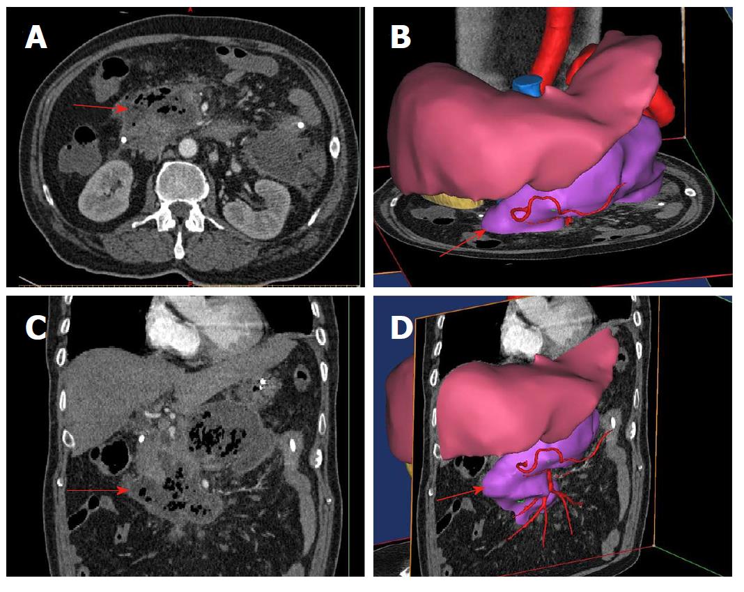
Figure 2 Cross-section, coronal-section and three-dimensional reconstruction images of the right-side puncture path for percutaneous catheter drainage in Patient No.
4. The red arrow represent the fictional direction and path of puncture.
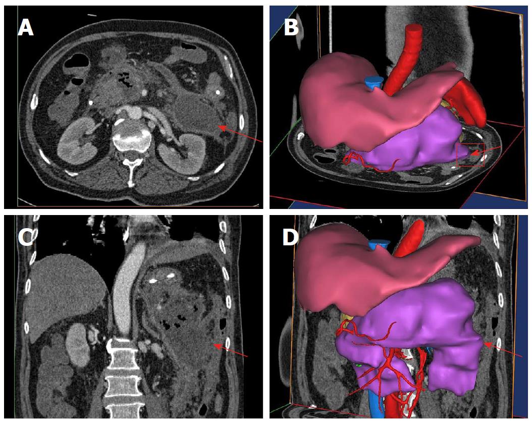
Figure 3 Cross-section, coronal section and three-dimensional reconstruction images of the upper left-side puncture path for percutaneous catheter drainage in Patient No.
4. The red arrow represents the fictional direction and path of puncture.
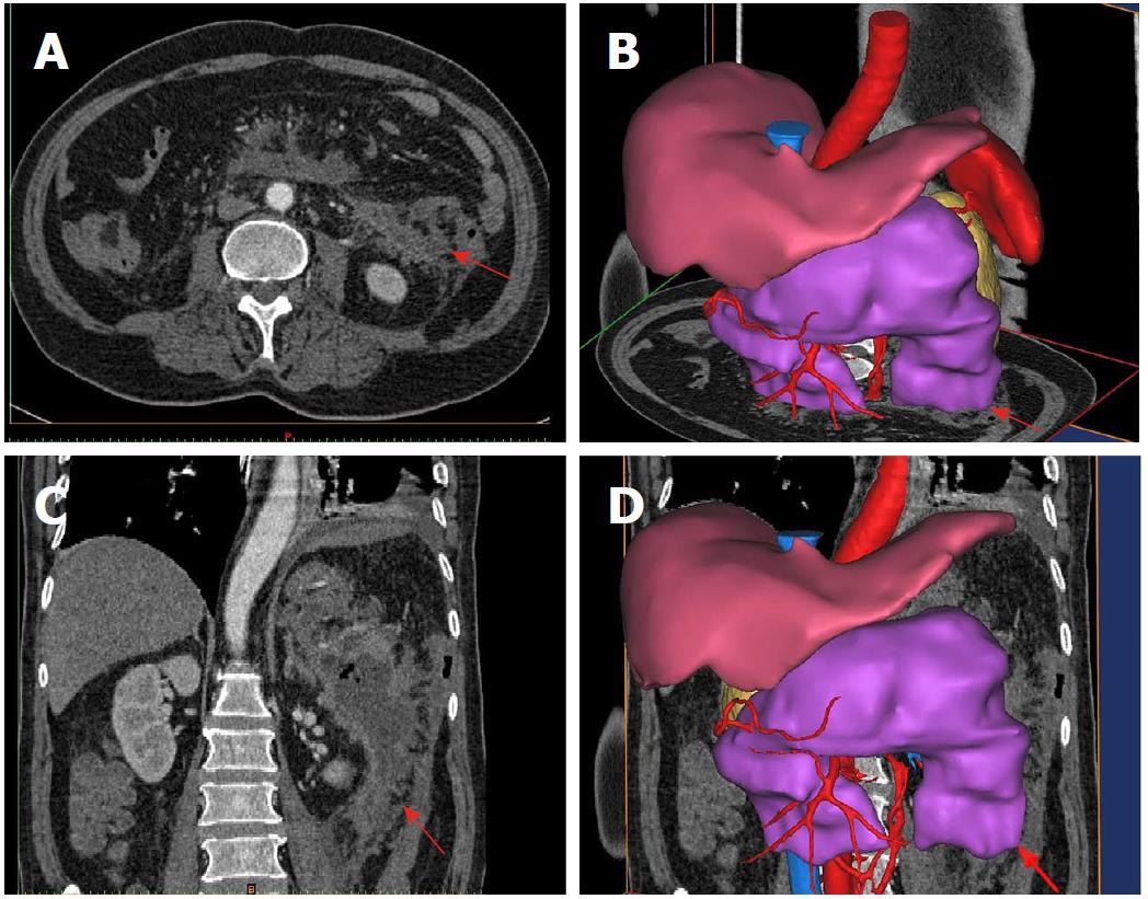
Figure 4 Cross-section, coronal section and three-dimensional reconstruction images of the lower left-side puncture path for percutaneous catheter drainage in Patient No.
4. The red arrow represents the fictional direction and path of puncture.
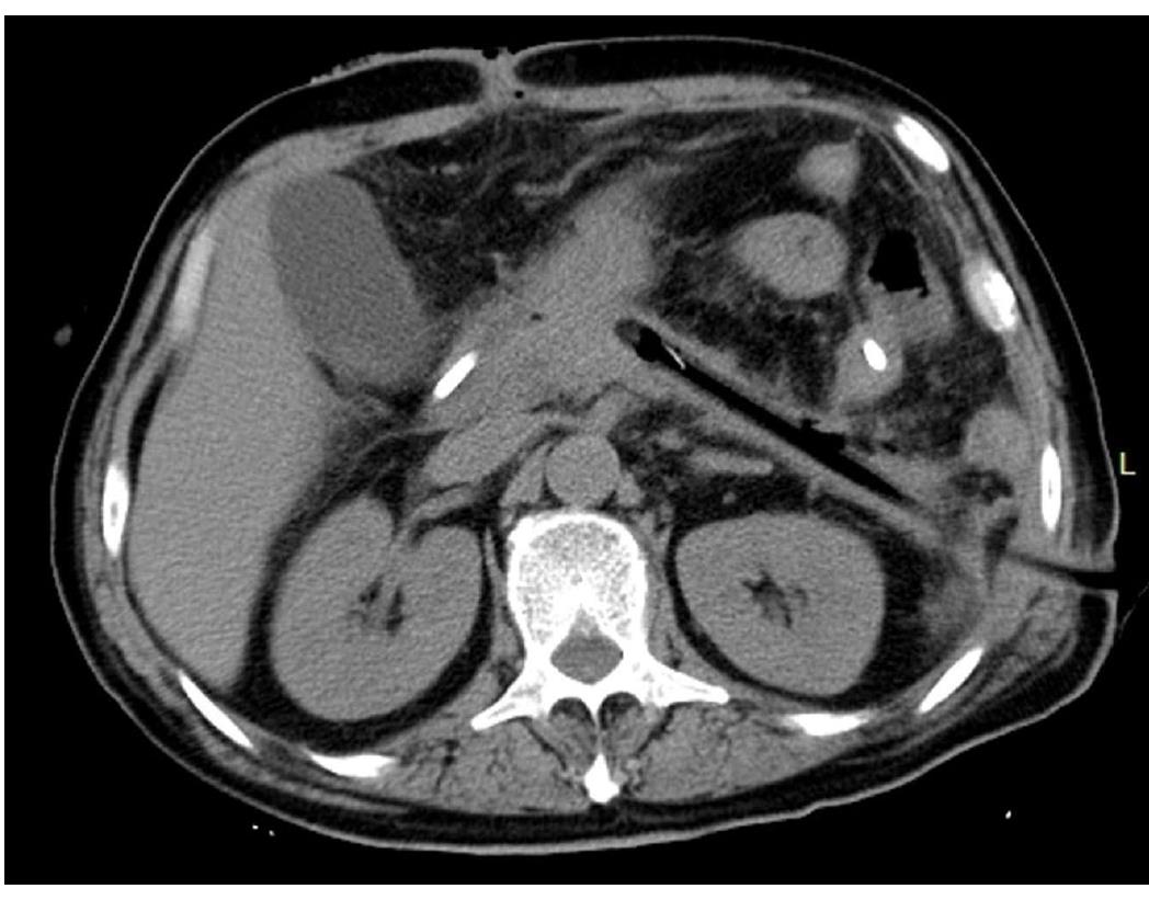
Figure 5 Image of abdominal CT reexamination of Patient No.
4. Abdominal CT reexamination 15 d after percutaneous nephroscopic necrosectomy showed that the abscess cavity disappeared and the drainage tube was unobstructed. CT: Computed tomography.
- Citation: Wang PF, Liu ZW, Cai SW, Su JJ, He L, Feng J, Xin XL, Lu SC. Usefulness of three-dimensional visualization technology in minimally invasive treatment for infected necrotizing pancreatitis. World J Gastroenterol 2018; 24(17): 1911-1918
- URL: https://www.wjgnet.com/1007-9327/full/v24/i17/1911.htm
- DOI: https://dx.doi.org/10.3748/wjg.v24.i17.1911


