Copyright
©The Author(s) 2017.
World J Gastroenterol. Mar 7, 2017; 23(9): 1645-1656
Published online Mar 7, 2017. doi: 10.3748/wjg.v23.i9.1645
Published online Mar 7, 2017. doi: 10.3748/wjg.v23.i9.1645
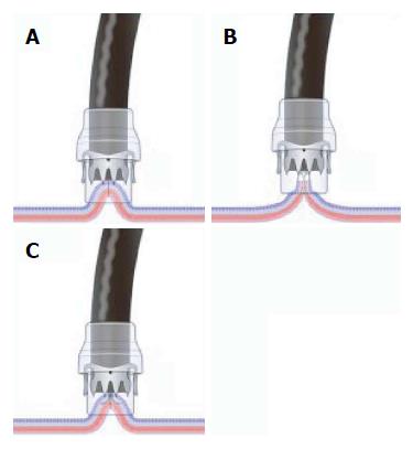
Figure 1 Key factor for the success of over-the-scope clips: schema of three suction methods into the application cap of the target lesion.
A: Simple suction, similar to endoscopic variceal band ligation (simple suction method); B: Assist of grasping forceps: Twin Grasper device (Twin Grasper method); C: Assist of tissue anchoring device (Anchor assist).
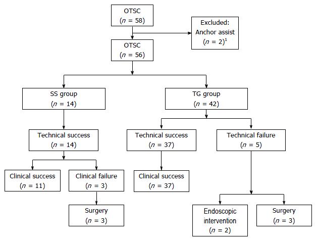
Figure 2 Flow diagram of patient enrollment and outcomes.
1Clinical outcomes of two cases that used the Anchor: one case with an incomplete closure of an esophageal-gastric anastomotic leakage, and another case with clinical success of gastric fistula after percutaneous endoscopic gastrostomy. SS: Simple suction; TG: Twin Grasper.
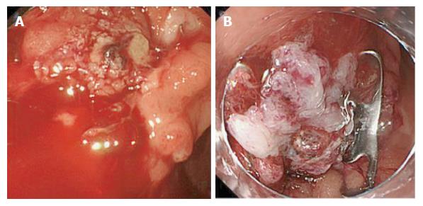
Figure 3 Representative clinical success case in the simple suction-group that exhibited refractory bleeding with a defect size of ≤ 10 mm and an immediate duration since onset.
A: A spurting, bleeding ulcer that was located in a rectal anastomotic site; B: Complete hemostasis via over-the-scope clip closure using the simple suction method after the failure of conventional endoscopic intervention.
Figure 4 Representative clinical success case in the Twin Grasper-group of a leak with a defect size of ≤ 10 mm and an immediate duration since onset.
A: An iatrogenic perforation site approximately 10 mm in size, located in the 2nd portion of the duodenum during endoscopic retrograde cholangiopancreatography; B: Application of the Twin Grasper device; C: Complete defect closure three months after over-the-scope clip deployment.
Figure 5 Representative case with an over-the-scope clips complication.
A: A delayed perforation with a 50-mm defect size after gastric endoscopic submucosal dissection; B: The misplacement of the over-the-scope clips to the exposed muscularis propria induced additional tears (black arrows); C: The defect could not be closed by the Twin Grasper due to a narrow lumen located in the prepylorus.
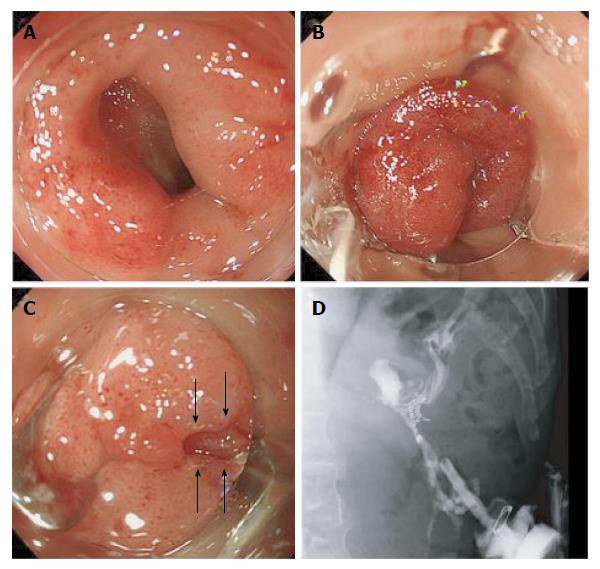
Figure 6 Clinically unsuccessful case in the simple suction-group of a fistula with a defect size of ≤ 10 mm and a chronic duration.
A: An 8-mm gastric fistula after interventional endoscopic ultrasound for a pseudo-pancreatic cyst; B: Complete closure with one over-the-scope clip (OTSC) using the simple suction method; C: A delayed leakage (black arrows) that occurred 2 wk after OTSC placement; D: X-ray contrast photo via a percutaneous drainage tube that revealed the delayed leakage.
- Citation: Kobara H, Mori H, Fujihara S, Nishiyama N, Chiyo T, Yamada T, Fujiwara M, Okano K, Suzuki Y, Murota M, Ikeda Y, Oryu M, AboEllail M, Masaki T. Outcomes of gastrointestinal defect closure with an over-the-scope clip system in a multicenter experience: An analysis of a successful suction method. World J Gastroenterol 2017; 23(9): 1645-1656
- URL: https://www.wjgnet.com/1007-9327/full/v23/i9/1645.htm
- DOI: https://dx.doi.org/10.3748/wjg.v23.i9.1645


