Copyright
©The Author(s) 2017.
World J Gastroenterol. Feb 28, 2017; 23(8): 1507-1512
Published online Feb 28, 2017. doi: 10.3748/wjg.v23.i8.1507
Published online Feb 28, 2017. doi: 10.3748/wjg.v23.i8.1507
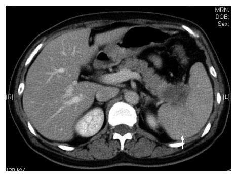
Figure 1 The low density tumor in the pancreatic tail (white arrow).
Figure 2 Fluorodeoxyglucose-positron emission tomography scan showing the swollen part in the pancreas tail and umbilical region (white arrow) with abnormal accumulation.
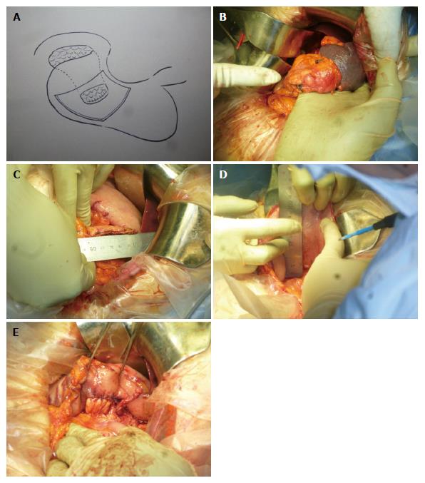
Figure 3 Our procedure.
A: We invaginate the pancreatic stump into the posterior wall of the stomach with single layer anastomosis using a 3-0 absorbable suture; B: The pancreatic tail tumor is indicated by the index finger on the left-hand side; C: Measurement of the width of the stump; D: The cutting length is determined at about 80% of the stump width; E: Anastomosis is completed. The pancreatic stump is invaginated into the posterior wall of the stomach.
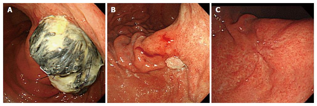
Figure 4 The change of the invaginated pancreatic stump in the stomach.
A: The invaginated pancreatic stump in the stomach after 1 wk; B: The stump after 3 mo; C: The stump after 1 year.
- Citation: Katsura N, Kawai Y, Gomi T, Okumura K, Hoashi T, Fukuda S, Takebayashi K, Shimizu K, Satoh M. Preventing pancreatic fistula after distal pancreatectomy: An invagination method. World J Gastroenterol 2017; 23(8): 1507-1512
- URL: https://www.wjgnet.com/1007-9327/full/v23/i8/1507.htm
- DOI: https://dx.doi.org/10.3748/wjg.v23.i8.1507


