Copyright
©The Author(s) 2017.
World J Gastroenterol. Feb 14, 2017; 23(6): 1113-1118
Published online Feb 14, 2017. doi: 10.3748/wjg.v23.i6.1113
Published online Feb 14, 2017. doi: 10.3748/wjg.v23.i6.1113
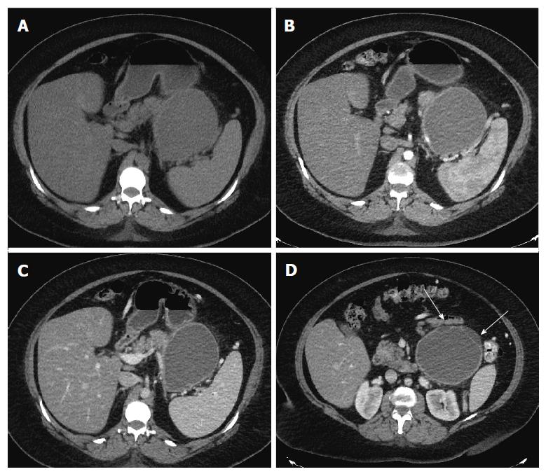
Figure 1 Axial computed tomography images.
Axial computed tomography (CT) images in unenhanced (A), pancreatic (B) and portal venous phase (C) show a well circumscribed, thin walled, large fluid density cystic lesion arising from and replacing the pancreatic body and tail, which abuts the spleen. The lesion does not exhibit post-contrast enhancement. Axial CT image at a lower level in portal venous phase (D) shows several thin septations and small loculations (arrows).
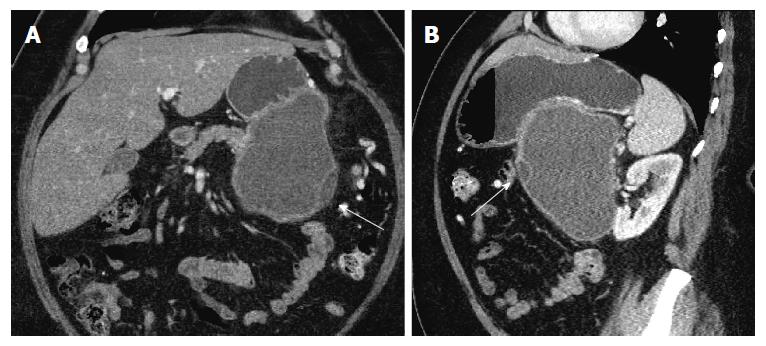
Figure 2 Coronal (A) and sagittal (B) images in portal venous phase show the same lesion abutting the stomach in its superior aspect.
Thin septations and small loculations are again noted (arrows).
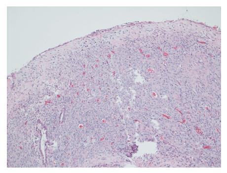
Figure 3 Cyst wall lined by bland cubo-columnar epithelium resting on a layer of cellular spindle cell stroma with thin walled blood vessels.
Hematoxylin and eosin staining, magnification × 100.
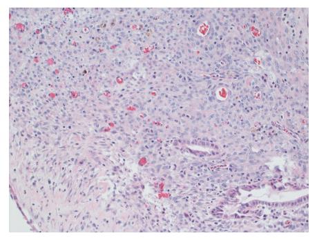
Figure 4 Cellular stroma shows bland spindle cells enclosing few benign glands and hemosiderin laden macrophages.
Hematoxylin and eosin staining, magnification × 200.
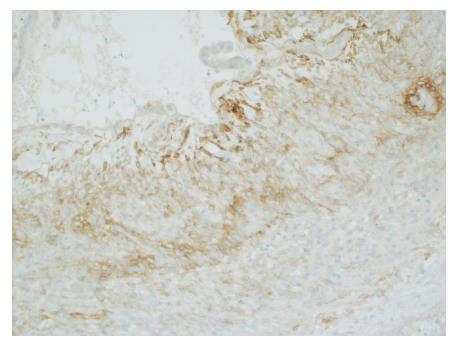
Figure 5 Positive staining for CD10 in the cellular spindloid stroma.
Immunohistochemical stain for CD10, magnification × 100.
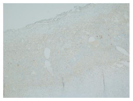
Figure 6 Negative staining for inhibin in the cellular spindloid stroma.
Immunohistochemical stain for inhibin, magnification × 100.
- Citation: Mederos MA, Villafañe N, Dhingra S, Farinas C, McElhany A, Fisher WE, Van Buren II G. Pancreatic endometrial cyst mimics mucinous cystic neoplasm of the pancreas. World J Gastroenterol 2017; 23(6): 1113-1118
- URL: https://www.wjgnet.com/1007-9327/full/v23/i6/1113.htm
- DOI: https://dx.doi.org/10.3748/wjg.v23.i6.1113


