Copyright
©The Author(s) 2017.
World J Gastroenterol. Feb 7, 2017; 23(5): 919-925
Published online Feb 7, 2017. doi: 10.3748/wjg.v23.i5.919
Published online Feb 7, 2017. doi: 10.3748/wjg.v23.i5.919
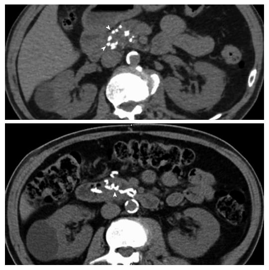
Figure 1 Computerized tomography scan without contrast showing calcifications in the pancreaticoduodenal arcade.
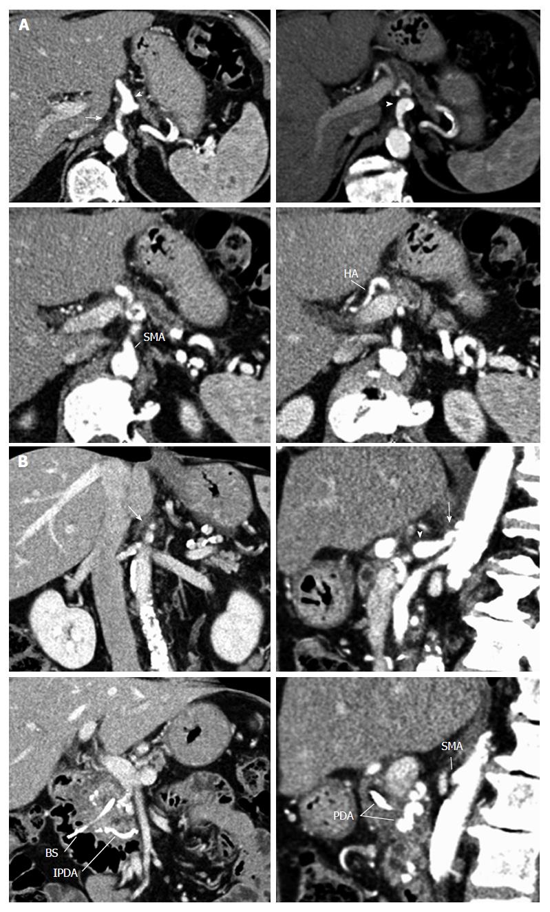
Figure 2 Features of median arcuate ligament.
Arterial phase of the axial computerized tomography (CT) scan (A), along with coronal and sagittal CT-scan reconstructions (B) showing severe stenosis of the celiac trunk from extrinsic compression by dense fibrous tissue (arrow) and poststenotic dilation of the proximal celiac trunk (arrowhead). SMA: Superior mesenteric artery; HA: Hepatic artery; PDA: Pancreaticoduodenal arcade; IPDA: Inferior pancreaticoduodenal artery; BS: Biliary stent.
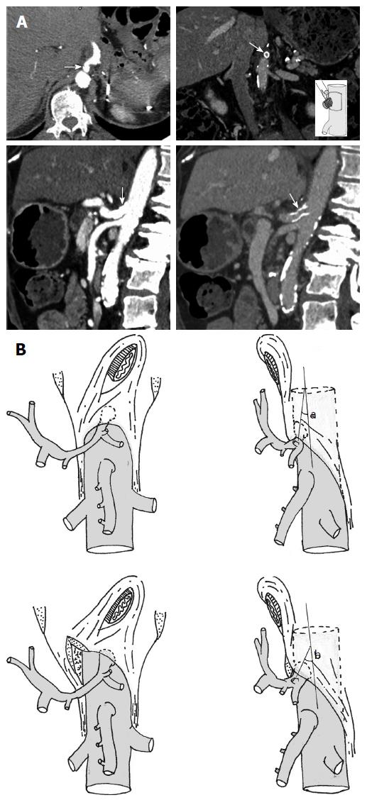
Figure 3 Computerized tomography scan.
Computerized tomography scan after stenting of the celiac trunk (A), drawings showing the angle between the aorta and celiac trunk before and after median arcuate ligament release (B).
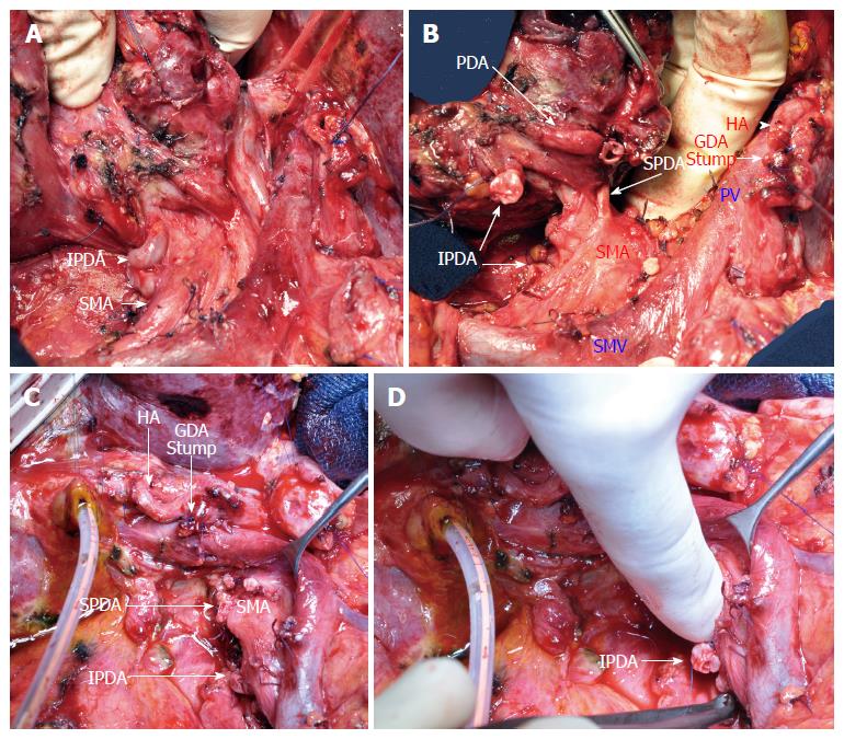
Figure 4 Pancreaticoduodenectomy.
Operative views (A, B) after mobilization of the specimen showing a very large inferior pancreaticoduodenal artery (IPDA) and the pancreaticoduodenal arcade. After resection (C, D), the stumps of the pancreaticoduodenal arteries (superior pancreaticoduodenal artery and IPDA) are shown. SMA: Superior mesenteric artery; HA: Hepatic artery; GDA: Gastroduodenal artery; SMV: Superior mesenteric vein; PV: Portal vein.
- Citation: Guilbaud T, Ewald J, Turrini O, Delpero JR. Pancreaticoduodenectomy: Secondary stenting of the celiac trunk after inefficient median arcuate ligament release and reoperation as an alternative to simultaneous hepatic artery reconstruction. World J Gastroenterol 2017; 23(5): 919-925
- URL: https://www.wjgnet.com/1007-9327/full/v23/i5/919.htm
- DOI: https://dx.doi.org/10.3748/wjg.v23.i5.919


