Copyright
©The Author(s) 2017.
World J Gastroenterol. Dec 28, 2017; 23(48): 8465-8488
Published online Dec 28, 2017. doi: 10.3748/wjg.v23.i48.8465
Published online Dec 28, 2017. doi: 10.3748/wjg.v23.i48.8465
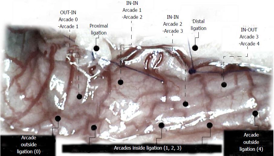
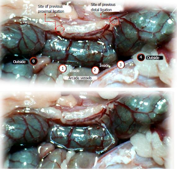
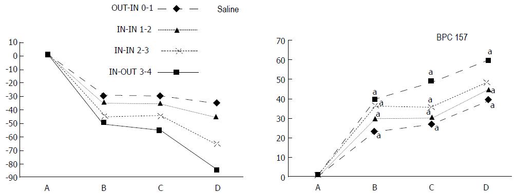
Figure 1 Assessment of arcade vessels arcade 0; arcade 1, 2, 3; arcade 4 (dorsal colon side before the initiation of therapy, USB microscope camera).
The ligations include three major arcades within the ligations (arcade 1, 2, 3), and thereby the possibility of bypassing both obstructions caused by the proximal and distal ligations (full lines) by an additional network of vessels located between the arcades (outside (arcade 0, arcade 4) and inside ligation (arcade 1, 2, 3); outside (arcade 0)-inside arcade 1); inside -inside(arcade 1-arcade 2; arcade 2-arcade 3); inside (arcade 3)-outside (arcade 4) (dashed lines) at the both ventral and dorsal sides of the colon. Note that there is no contact between inside arcade 3 and outside arcade 4. The appearance of the arcade vessels at specific time points [A (1 minute before therapy), B (next 5 min), C (the next 5 minutes, until the 10th min), D (until the end of the 15th min)] was assessed between (1) the last arcade proximal to first ligation (arcade 0) at the first arcade distal to first ligation, inside the ligated segment (arcade 1) (OUT-IN) (0-1, Figure 2, ♦--, Figures 5, 6, 13, 14); (2) between the next arcade (arcade 2), middle arcade inside ligations (IN-IN) (1-2, Figure 2; ▲, Figures 5, 6, 13, 14) and the following arcade (2-3, Figure 2; --X--, Figures 5, 6, 13, 14), and finally presentation to the first outside arcade distal to the second ligation (arcade 4) (IN-OUT) (3-4, Figure 2; ■, Figures 5, 6, 13, 14).
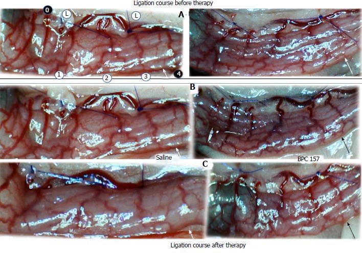
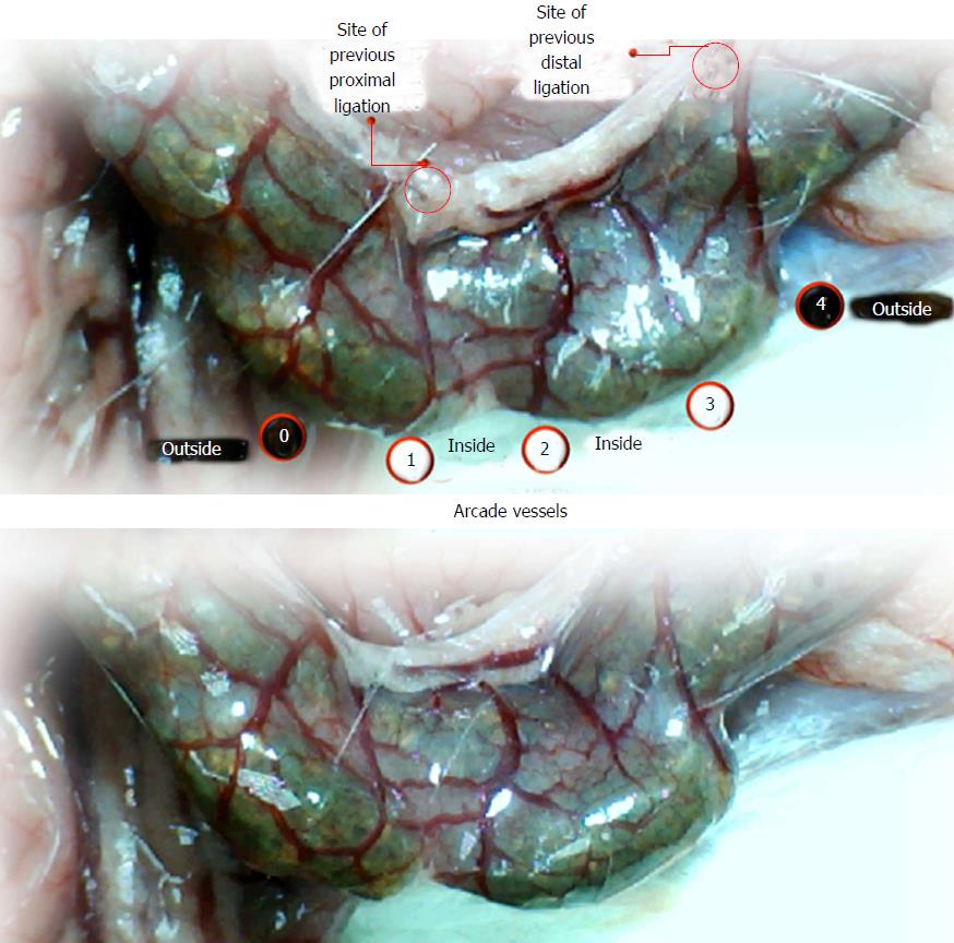
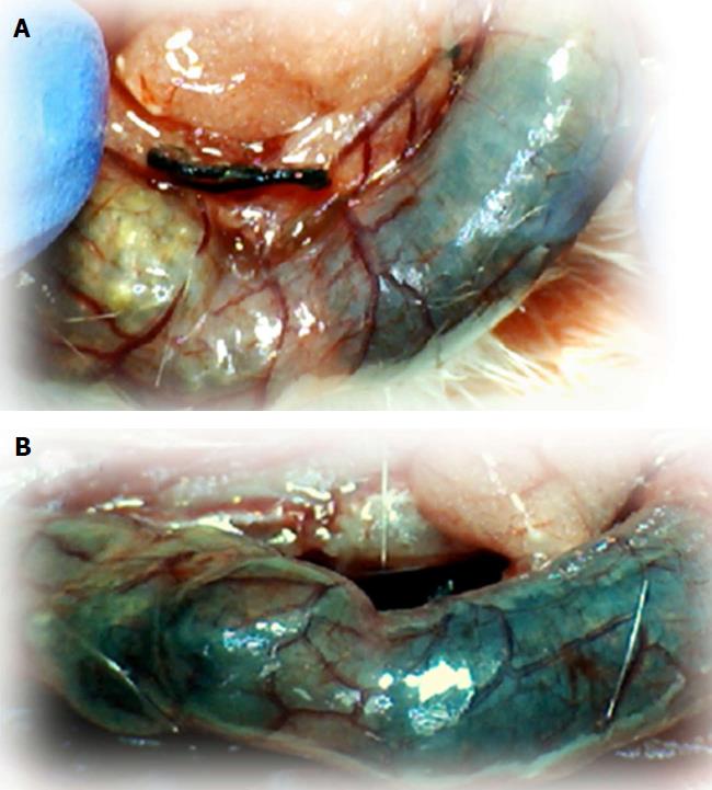
Figure 2 Progressive disappearance (A, saline bath; B, C, left) or recovery (BPC 157 bath; B, C, right) of blood vessels after ligation of the dorsal colon of IC rats (USB microscope camera).
Ligation course before therapy (A); ligation course after therapy (B, C). A. Left colic artery and vein major arcade vessels obstructed by two ligations, proximal (left) and distal (right) (L), arcade vessels outside 0, 4 (black circles) or inside 1, 2, 3 (white circles), inside-outside connection absent (arcade 3-arcade 4) (white arrows). B. Five minutes later: inside-outside connection still absent (saline bath, left) (white arrow) or reestablished (black arrow); vessel presentation markedly increased (BPC 157 bath, right). C. From the 10th minute through the end of the 15th minute. Progressive vessel disappearance (saline bath, left) (white arrow); recovered blood vessels (BPC 157 bath, right) (black arrow).
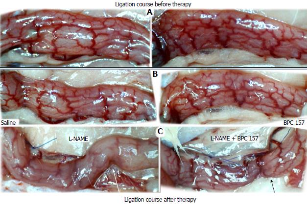
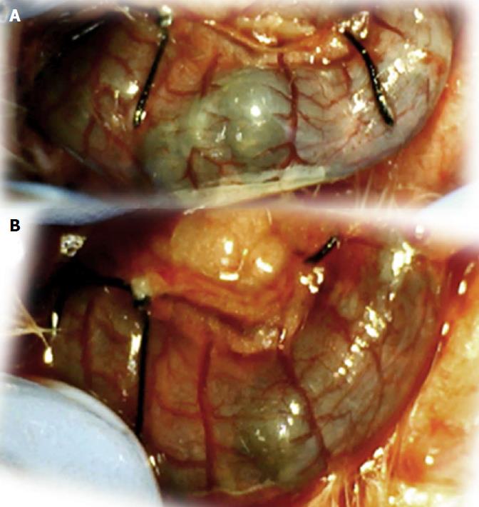
Figure 3 Blood vessels after ligation in IC rats (ventral colon side of the colon, before application of medication).
A: Progressive disappearance of blood vessels (saline bath, B, ventral colon side; L-NAME, C, dorsal colon side, left) or recovery (BPC 157 bath, B, ventral colon side; L-NAME + BPC 157, C, dorsal colon side, right). Ligation course before therapy (A); ligation course after therapy (B, C). A. Ventral side of the colon showing the presence of an abundant collateral network with inside-outside contact, unlike the relatively scarce arcade collateral network and absent inside-outside contact on the dorsal side (see Figure 2). B. Five minutes later, the number of vessels on the ventral side of the colon is markedly decreased; the inside-outside connection is still present (saline bath, left); or vessel presentation is markedly increased (BPC 157 bath, right). C. From the 10th minute through the end of the 15th minute. Dorsal side of the colon showing progressive vessel disappearance, inside-outside contact largely absent (L-NAME bath, left) (white arrow), unlike recovered blood vessels and inside-outside contact (L-NAME+BPC 157 bath, right (black arrow).
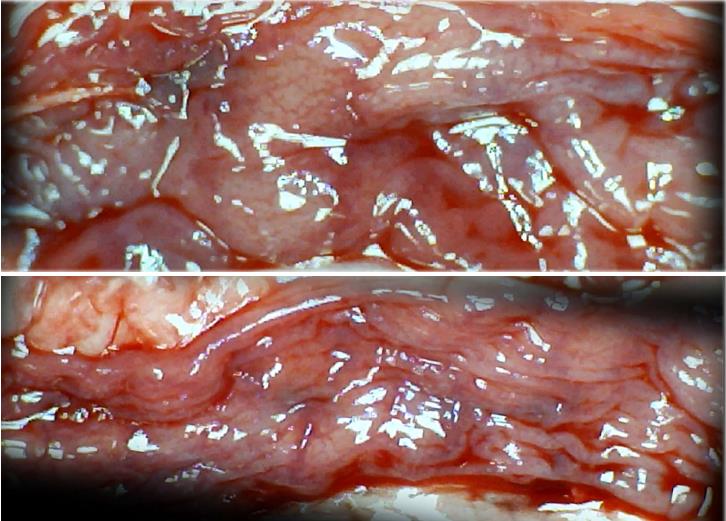
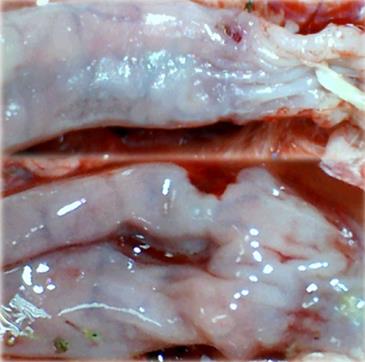
Figure 4 Characteristic appearance of the colon in IC rats 15 min after ligation, upon colon opening before sacrifice; USB microscope camera.
Extremely large pale areas without mucosal folds were observed after saline bath treatment (upper), in contrast to the preserved mucosal folds seen after BPC 157 bath treatment (lower) in rats that underwent obstruction of the left colic artery and vein for 15 min. The animals initially received bath medication consisting of saline (upper) or BPC 157 (lower).
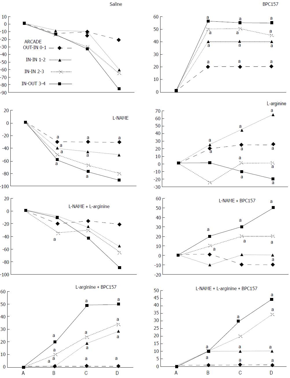
Figure 5 IC rats.
Per cent of vessels present between arcade vessels next to the proximal (arcade 0) and distal (arcade 4) ligatures and within the ligated area (arcade 1, 2, 3) on the dorsal side of the colon at 15 min following therapy (as 100%); mean ± SD. The gross appearance of the tissue was recorded using a USB microscope camera. The following time points were assessed: A - after ligation and before therapy (1 min); B - 5 min after the application of medication; C - between 5 and 10 min after the application of medication; D - from 10 min after the application of medication until the end of the observation at 15 min. At 1 min post-injury, medication (/kg, 1 ml bath/rat) consisting of BPC 157 (10 µg), the NOS blocker L-NAME (5 mg) and the NOS substrate L-arginine (100 mg) alone or combined, or an equal volume of a saline bath (controls) was applied to the 25-mm blood-flow-deprived colon segment; the rats were sacrificed at 15 minutes. For clarity, the SD is not shown on the graph; the SD was never higher than 10% of the mean. aP < 0.05 at least vs control.
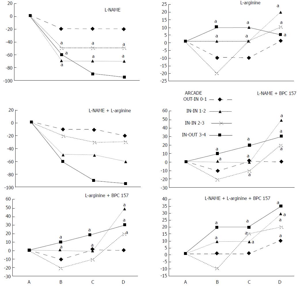
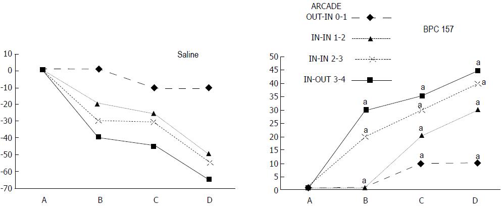
Figure 6 IC rats.
Per cent of vessels present between arcade vessels, next to the proximal ligature (arcade 0), next to the distal ligature (arcade 4), and within the ligated area (arcade 1, 2, 3), on the ventral side of the colon at 15 min following therapy (as 100%); mean ± SD. The gross appearance of the tissue was recorded using a USB microscope camera. The following time points were assessed: A - after ligation and before therapy (1 min); B - 5 min after the application of medication; C - between 5 and 10 min after the application of medication; D - from 10 min after the application of medication until the end of the observation at 15 min. At 1 min post-injury, medication (/kg, 1 mL bath/rat) consisting of BPC 157 (10 µg), the NOS blocker L-NAME (5 mg) and the NOS substrate L-arginine (100 mg), alone or combined, or an equal volume of a saline bath (controls) was applied to the 25-mm blood-flow-deprived colon segment; the rats were sacrificed at 15 min. For clarity, the SD is not shown on the graph; the SD was never higher than 10% of the mean. aP < 0.05 at least vs control.
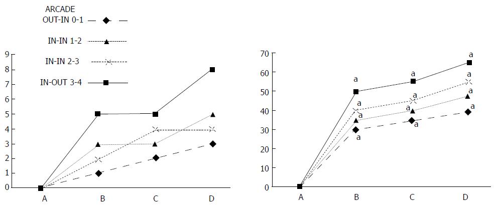
Figure 7 IC + RL rats.
Per cent of vessel presentation between arcade vessels, next to the previous proximal ligature (arcade 0), next to the previous distal ligature (arcade 4), and within the previous ligated area (arcade 1, 2, 3) on the dorsal side of the colon at 15 min after therapy (as 100%); mean ± SD. The gross appearance of the tissue was recorded using a USB microscope camera. The following time points were assessed: A: After ligation and before therapy (1 min); B: 5 min after the application of medication; C: Between 5 and 10 min after the application of medication; D: From 10 min after the application of medication until the end of the observation at 15 min. At 1 min post-injury, BPC 157 (10 μg /kg, 1 mL bath/rat) or an equal volume of a saline bath (controls) was applied to the reperfused 25-mm colon segment; the rats were sacrificed at 15 min. For clarity, the SD is not shown on the graph; the SD was never higher than 10% of the mean. aP < 0.05 at least vs control.
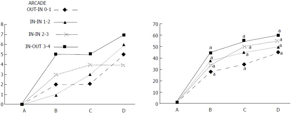
Figure 8 IC + RL rats.
Per cent of vessels present between arcade vessels, next to the previous proximal ligature (arcade 0), next to the previous distal ligature (arcade 4), and within the previous ligation (arcade 1, 2, 3) on the ventral side of the colon 15 min following therapy (as 100%); mean ± SD. The gross appearance of the tissue was recorded using a USB microscope camera. The following time points were assessed: A: After ligation and before therapy (1 min); B: 5 min after the application of medication; C: Between 5 and 10 min after the application of medication; D: From 10 min after the application of medication until the end of the observation at 15 min. At 1 min post-injury, medication (BPC 157, 10 μg/kg, 1 mL bath/rat) or an equal volume of a saline bath was applied to the reperfused 25-mm colon segment; the rats were sacrificed 15 min later. For clarity, the SD is not shown on the graph; the SD was never higher than 10% of the mean. aP < 0.05 at least vs control.
Figure 9 IC + RL rats (controls).
Vessel presentation between arcade vessels next to the previous proximal ligature (arcade 0), next to the previous distal ligature (arcade 4), and within the previous ligation (arcade 1, 2, 3) on the dorsal side of the colon at 1 min reperfusion time and prior to saline bath application (upper). Similar vessel presentation is apparent immediately after application of a saline bath (1 mL bath/rat) (controls) to the reperfused 25-mm colon segment (lower).
Figure 10 IC + RL rats (BPC 157-treated rats).
Vessel presentation between arcade vessels, next to the previous proximal ligature (arcade 0), next to the previous distal ligature (arcade 4), and within the previous ligation (arcade 1, 2, 3) on the dorsal side of the colon at 1 minute reperfusion time and before saline bath application (upper). Increasing vessel presentation forming “honeycomb” structures between arcades can be seen immediately after BPC 157 treatment (BPC 157 (10 μg/kg), 2 μg/mL bath/rat) of the reperfused 25-mm colon segment (lower).
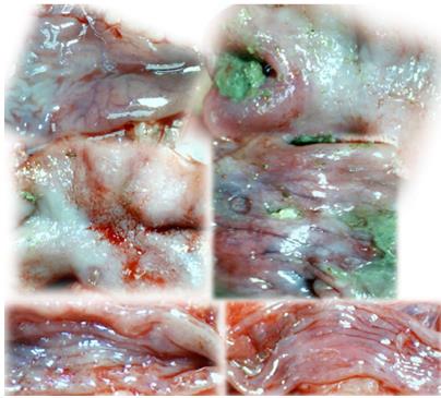
Figure 11 IC + RL rats (controls) at 15 min reperfusion time.
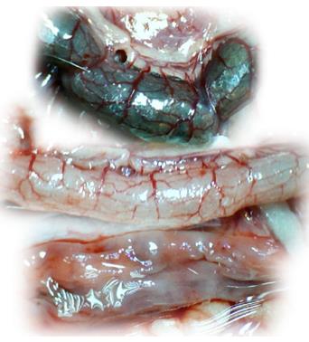
At 1 min reperfusion time, saline (1 mL bath/rat) was applied to the reperfused 25-mm colon segment (lower). Relatively poor vessel presentation between arcade vessels is apparent in the full colon segment (upper); colon segment cleaned with water (middle); progressively appearing, extremely large pale areas without mucosal folds upon colon opening before sacrifice (lower).
Figure 12 IC + RL rats (BPC 157-treated rats) at 15 min reperfusion time.
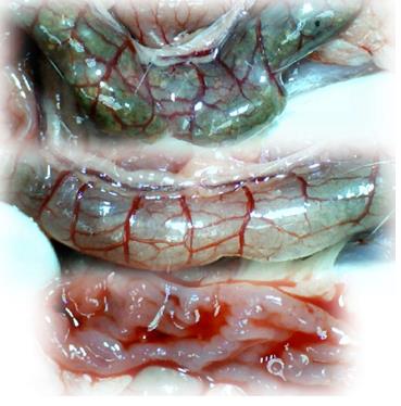
At 1 min reperfusion time, BPC 157 (10 μg/kg, 2 µg/mL bath/rat) was applied to the reperfused 25-mm colon segment (lower). Increasing vessel presentation between arcade vessels is apparent in the full colon segment (upper); colon segment cleaned with water (middle); fully recovered mucosa and mucosal folds upon colon opening before sacrifice (lower).
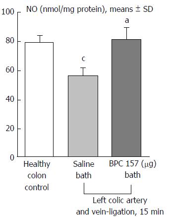
Figure 13 NO levels in the colon tissue of IC rats at 15 min ligation time determined using the Griess reaction.
BPC 157 (10 μg/kg, 1 mL bath/rat) or an equal volume of a saline bath (controls) was applied to the 25-mm blood-flow-deprived colon segment. aP < 0.05 vs saline; cP < 0.05 vs healthy colon.
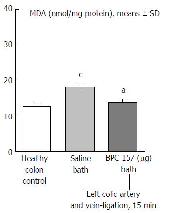
Figure 14 Malondialdehyde levels in the colon tissue of IC rats at 15 min ligation time, determined by quantifying thiobarbituric acid reactivity as malondialdehyde equivalents.
BPC 157 (10 μg/kg, 1 ml bath/rat) or an equal volume of a saline bath (controls) was applied to the 25-mm blood-flow-deprived colon segment. aP < 0.05 vs saline; cP < 0.05 vs healthy colon.
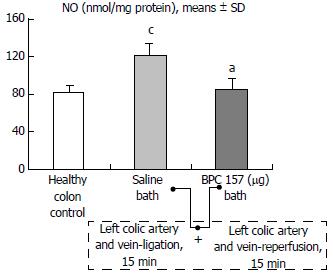
Figure 15 NO levels in the colon tissue of IC + RL rats after 15 min of reperfusion, determined using the Griess reaction.
At 1 min reperfusion time, BPC 157 (10 μg/kg, 2 μg/1 mL bath/rat) or an equal volume of a saline bath (controls) was applied to the reperfused 25-mm colon segment. aP < 0.05 vs saline; cP < 0.05 vs healthy colon.
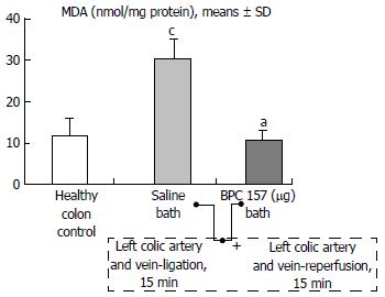
Figure 16 Malondialdehyde levels in the colon tissue of IC + RL rats after 15 min of reperfusion, determined by quantifying thiobarbituric acid reactivity as malondialdehyde equivalents.
At 1 min reperfusion time, BPC 157 (10 μg/kg, 2 μg/1 mL bath/rat) or an equal volume of a saline bath (controls) was applied to the reperfused 25-mm colon segment. aP < 0.05 vs saline; cP < 0.05 vs healthy colon.
Figure 17 IC + OB rats that underwent additional colon obstruction for three days presented severely impaired gross arcade vessels immediately after the additional colon obstruction was removed (A, before therapy, upper); after saline bath application, they exhibited an increasing disappearance of arcade vessels, particularly on the dorsal side of the colon (B, 5 min after therapy, lower).
The images were obtained using a USB microscope camera.
Figure 18 IC + OB rats that underwent additional colon obstruction for the first three days presented severely impaired gross arcade vessels immediately after the additional colon obstruction was removed (A, before therapy, upper); by contrast, after bath treatment with BPC 157, all of the arcade vessels vigorously reappeared, together with interconnecting collaterals between the arcades.
Rapid recovery of blood flow also occurred, and this was especially evident on the dorsal side of the colon (B, 5 min after therapy, lower). The images were obtained using a USB microscope camera.
Figure 19 In IC + OB rats that underwent additional colon obstruction for three days, colon opening revealed pale flattened areas without mucosal folds immediately after additional colon obstruction was removed.
The images were obtained using a USB microscope camera.
Figure 20 IC + OB rats underwent additional colon obstruction for three days; the colon was then opened before sacrifice on day 10 [i.
e., one week after the additional colon obstruction had been removed and bath therapy applied (saline, upper and middle; BPC 157, lower)]. The examination revealed progressive worsening in the saline-treated animals (upper and middle) and recovery in the BPC 157-treated animals (lower) (see Figure 11). The control rats exhibited extremely large pale areas without mucosal folds covering the entire area between ligations (middle); such areas were present even beyond the area of the ligation (upper), together with ulcerations in the deprived, apparently enlarged colon segment (middle). BPC 157-treated rats exhibited almost completely spared mucosa (very small pale areas) and no ulceration; the previously ligated colon segment was of normal diameter (lower). The images were obtained using a USB microscope camera.
Figure 21 IC + OB rats.
Per cent of vessels present between arcade vessels, next to the proximal ligature (arcade 0), next to the distal ligature (arcade 4), and within the ligation (arcade 1, 2, 3) on the dorsal side of the colon at 15 min after therapy (as 100%); mean ± SD. The gross presentation was recorded using a USB microscope camera. After additional colon obstruction for three days, BPC 157 (10 μg/kg, 1 mL bath/rat) or an equal volume of a saline bath was applied to the 25-mm blood-flow-deprived colon segment 1 min after the colon obstruction was removed. The rats were sacrificed 15 min later, and the following time points were assessed: A: after ligation and before therapy (1 min); B: 5 min after the application of medication; C: between 5 and 10 min after the application of medication; D: from 10 min after the application of medication until the end of the observation at 15 min. For clarity, the SD is not shown on the graph; the SD was never higher than 10% of the mean. aP < 0.05 at least vs control.
Figure 22 IC + OB rats.
Per cent of vessels present between arcade vessels, next to the proximal ligature (arcade 0), next to the distal ligature (arcade 4), and within the ligated area (arcade 1, 2, 3) on the ventral side of the colon 15 min after therapy (as 100%); mean ± SD. The gross presentation was recorded using a USB microscope camera. After additional colon obstruction for three days, BPC 157 (10 μg/kg, 1 mL bath/rat) or an equal volume of a saline bath was applied to the 25-mm blood-flow-deprived colon segment 1 min after the colon obstruction was removed. The rats were sacrificed 15 min later, and the following time points were assessed: A - after ligation and before therapy (1 min); B - 5 min after the application of medication; C - between 5 and 10 min after the application of medication; D - from 10 min after the application of medication until the end of the observation at 15 min. For clarity, the SD is not shown on the graph; the SD was never higher than 10% of the mean. aP < 0.05 at least vs control.
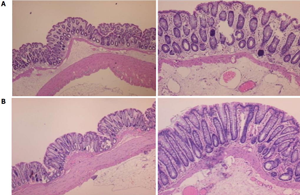
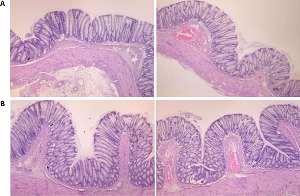
Figure 23 Characteristic microscopic appearance of the colon in IC (A, control) rats and BP 157-treated IC rats (B) 15 min after ligation.
HE, 4 × objective (left) and 10 × objective (right). A: Severe edema of the lamina propria and continuous diffuse edema of the submucosa can be observed. Pronounced dilatation and stasis of the submucosal blood vessels is also present. B: Mild edema of the lamina propria and focally present mild-to-intermediate edema of the submucosa can be observed. Although stasis of the submucosal blood vessels is present, the dilatation of the veins appears to be less pronounced.
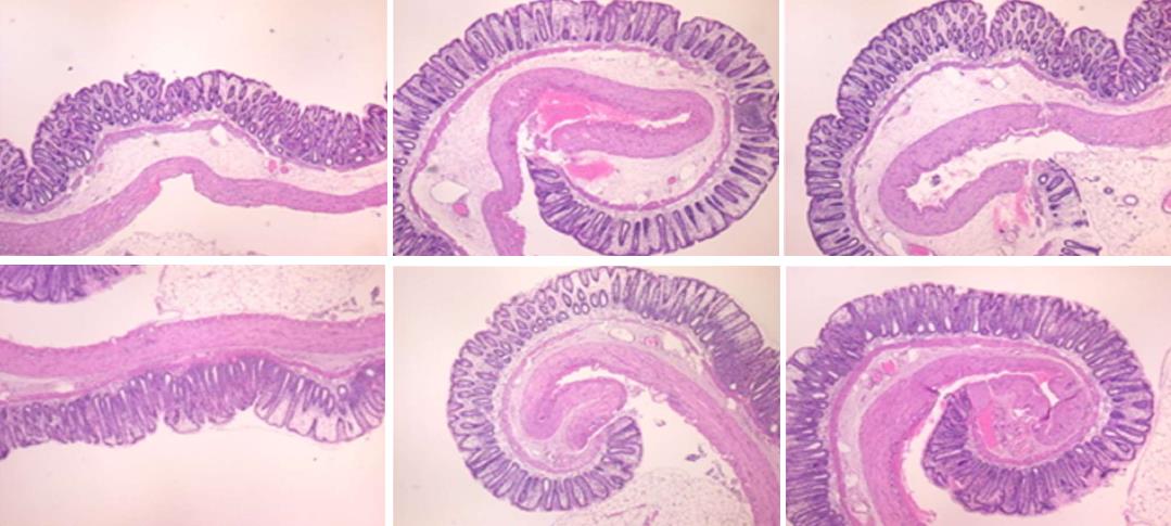
Figure 24 In IC + RL rats, 15 min of ligation followed by 15 min of full reperfusion period resulted in the appearance of even more mucosal and submucosal edema as well as more pronounced cyanosis (controls, upper, HE, 4 × objective).
This was consistently attenuated by BPC 157 treatment after the initiation of reperfusion (lower, HE, 4 × objective).
Figure 25 Microscopic appearance of the colon in IC + OB rats (A, control) and BPC 157-treated IC + OB rats (B) after additional colon obstruction for three days one week after the additional colon obstruction was removed.
Control (saline bath); 10 d; BPC 157, 10 d. HE, 4 × objective (left); 10 × objective (right). A: Mild edema of the lamina propria and diffuse mild-to-intermediate edema of the submucosa, along with the formation of collagen, can be observed. Stasis of the submucosal blood vessels is also present. The rugae are broadened and flattened, so the mucosa appears flattened macroscopically. B: Mild edema of the lamina propria and practically no edema of the submucosa is observed. Stasis of the submucosal blood vessels is also present but to a much lesser extent than in the controls. The rugae are histologically well formed and only occasionally slightly broadened. There is minimal formation of new collagen fibers in the submucosa.
- Citation: Duzel A, Vlainic J, Antunovic M, Malekinusic D, Vrdoljak B, Samara M, Gojkovic S, Krezic I, Vidovic T, Bilic Z, Knezevic M, Sever M, Lojo N, Kokot A, Kolovrat M, Drmic D, Vukojevic J, Kralj T, Kasnik K, Siroglavic M, Seiwerth S, Sikiric P. Stable gastric pentadecapeptide BPC 157 in the treatment of colitis and ischemia and reperfusion in rats: New insights. World J Gastroenterol 2017; 23(48): 8465-8488
- URL: https://www.wjgnet.com/1007-9327/full/v23/i48/8465.htm
- DOI: https://dx.doi.org/10.3748/wjg.v23.i48.8465


