Copyright
©The Author(s) 2017.
World J Gastroenterol. Dec 14, 2017; 23(46): 8227-8234
Published online Dec 14, 2017. doi: 10.3748/wjg.v23.i46.8227
Published online Dec 14, 2017. doi: 10.3748/wjg.v23.i46.8227
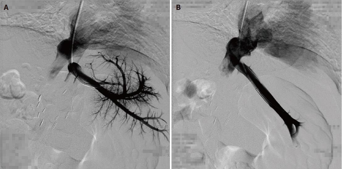
Figure 1 Left lobe graft for biliary atresia in a 2-year-old boy.
A: Hepatic venography shows severe blockage of the hepatic venous outflow. The pressure gradient between the right atrium and left vein was 13 mmHg. B: Hepatic venography after percutaneous transluminal venoplasty shows improved flow into the right atrium. The pressure gradient decreased to 3 mmHg.
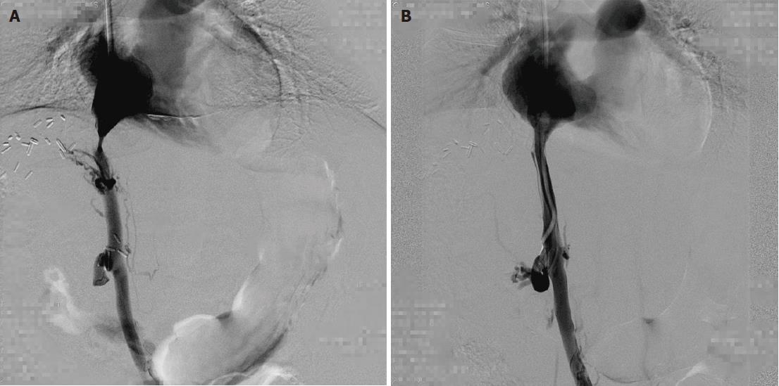
Figure 2 Left lobe graft for biliary atresia in a 10-month-old girl.
A: The inferior vena cava (IVC) image shows tight fibrotic stenosis at the superior anastomosis close to the right atrium. B: The IVC angiogram after angioplasty with a 6-20 mm balloon shows stricture resolution, no residual stenosis and free flow through the IVC into the right atrium.
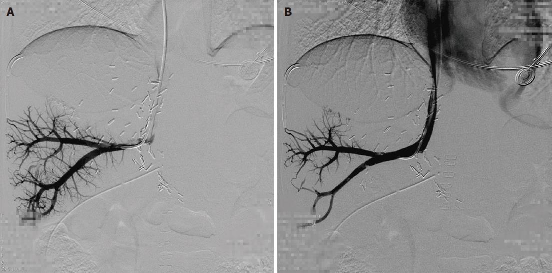
Figure 3 A 9-year-old male received cross-auxiliary double-domino donor liver transplantation.
One month after the initial procedure, stenosis recurred. A: Hepatic venography shows a stricture at the piggyback hepatic vein anastomosis. B: Hepatic vein angiography after balloon dilatation shows the disappearance of the stricture and free flow through the anastomosis.
- Citation: Zhang ZY, Jin L, Chen G, Su TH, Zhu ZJ, Sun LY, Wang ZC, Xiao GW. Balloon dilatation for treatment of hepatic venous outflow obstruction following pediatric liver transplantation. World J Gastroenterol 2017; 23(46): 8227-8234
- URL: https://www.wjgnet.com/1007-9327/full/v23/i46/8227.htm
- DOI: https://dx.doi.org/10.3748/wjg.v23.i46.8227


