Copyright
©The Author(s) 2017.
World J Gastroenterol. Nov 28, 2017; 23(44): 7840-7848
Published online Nov 28, 2017. doi: 10.3748/wjg.v23.i44.7840
Published online Nov 28, 2017. doi: 10.3748/wjg.v23.i44.7840
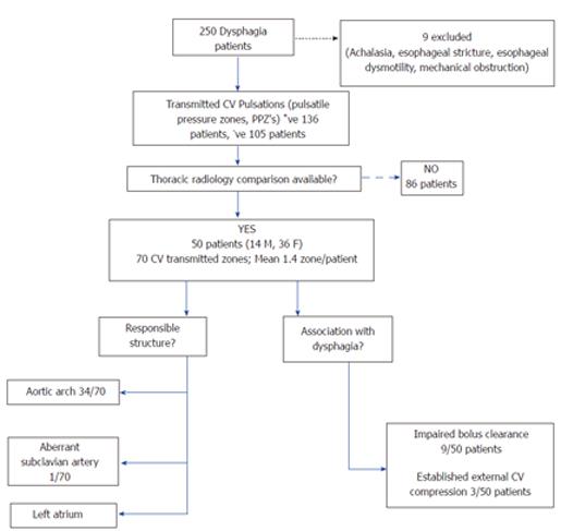
Figure 1 Selection of cases (dysphagia patients) with transmitted cardiovascular pulsations as noted on high resolution esophageal impedance manometry.
CV: Cardiovascular.
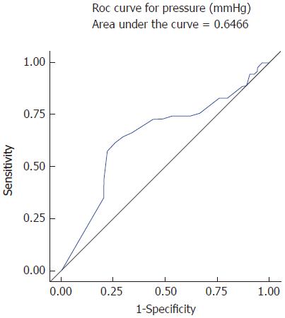
Figure 2 The receiver operator curve for pressure across pulsatile pressure zone with dysphagia shows area under the curve 0.
65 demonstrating weak significance. Hence even though pressure was a statistically significant variable on Wilcoxon Rank Sum analysis, it is a poor predictor of dysphagia caused by transmitted cardiovascular pulsations on esophagus by this model.
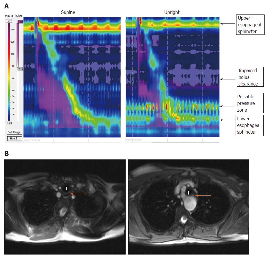
Figure 3 In the patient with dysphagia lusoria, bolus clearance was 10% in supine swallows and 90% upright swallows.
A: Pulsatile pressure zones (PPZ) on HREIM due to transmitted pulsations of aberrant subclavian artery and aortic arch in a patient with dysphagia lusoria, with impaired bolus clearance; B: Magnetic Resonance Angiogram of patient with dysphagia lusoria; LEFT patent esophagus (arrow) RIGHT esophageal compression secondary to aberrant subclavian artery (dysphagia lusoria); T: Trachea.
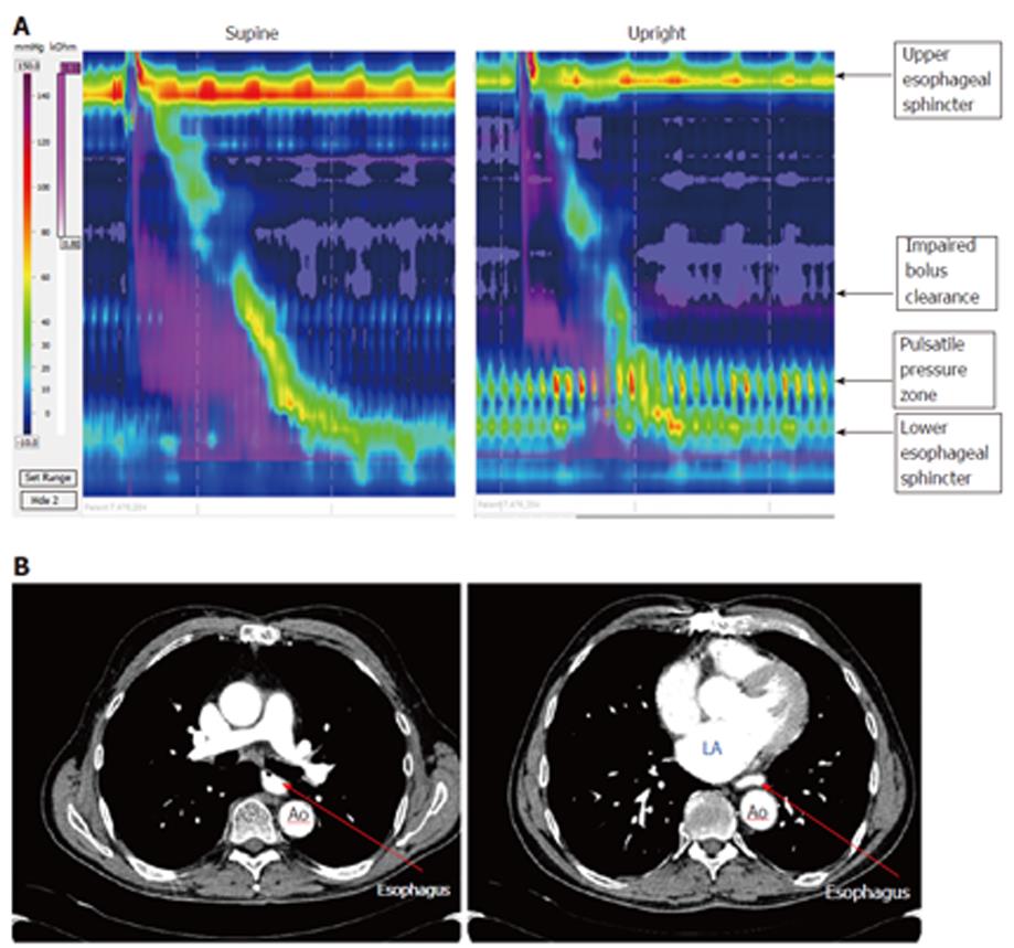
Figure 4 In one of the patients with enlarged left atrium, bolus clearance was 80% in supine swallows and 10 % upright.
A: Pulsatile pressure zone with impaired bolus clearance easily visible in a dysphagia patient in upright posture due to left atrial enlargement; B: Corresponding CT Scan Images of the same patient (Left) Esophagus patent in upper thorax (Right) Esophagus compressed between dilated left atrium (LA) and descending aorta (Ao).
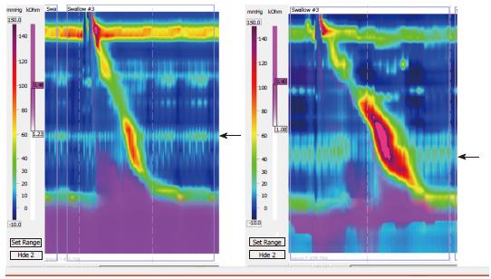
Figure 5 Pulsatile pressure zones observed in patients without dysphagia (see arrows).
- Citation: Chaudhry NA, Zahid K, Keihanian S, Dai Y, Zhang Q. Transmitted cardiovascular pulsations on high resolution esophageal impedance manometry, and their significance in dysphagia. World J Gastroenterol 2017; 23(44): 7840-7848
- URL: https://www.wjgnet.com/1007-9327/full/v23/i44/7840.htm
- DOI: https://dx.doi.org/10.3748/wjg.v23.i44.7840


