Copyright
©The Author(s) 2017.
World J Gastroenterol. Nov 14, 2017; 23(42): 7519-7530
Published online Nov 14, 2017. doi: 10.3748/wjg.v23.i42.7519
Published online Nov 14, 2017. doi: 10.3748/wjg.v23.i42.7519
Figure 1 Histological analyses of rat livers.
Representative photomicrographs of liver sections taken from sham-operated (A), mildly cholestatic (B) and severely cholestatic rats (C), stained with van Gieson to detect liver fibrosis (magnification × 5).
Figure 2 mRNA expression of CYP3A isoforms in rat livers.
Gene expression of CYP3A1 (A) and CYP3A2 (B). Results are mean ± SEM, obtained from 8 rats of each group. aP < 0.05, bP < 0.01 vs sham-operated rats; eP < 0.01 and fP < 0.001 vs rats with mild cholestasis.
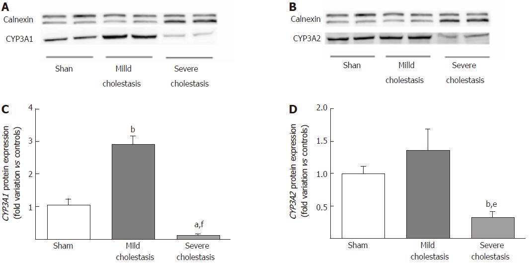
Figure 3 Microsomal protein expression of CYP3A isoforms in rat livers.
A representative Western blot experiment showing CYP3A1 (A) and CYP3A2 (B) protein expression in microsomal fractions obtained from sham-operated and cholestatic rats. Densitometric analysis of CYP3A1 (C) and CYP3A2 (D) protein bands. Results are mean ± SEM obtained from 8 rats of each group. Each experiment was conducted in triplicate. aP < 0.05, bP < 0.01 vs sham rats; eP < 0.01, fP < 0.001 vs mildly cholestatic rats.
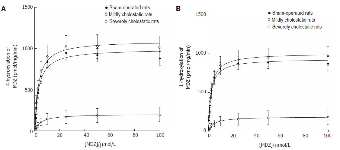
Figure 4 Enzymatic activity of CYP3A in microsomes obtained from sham-operated, mildly and severely cholestatic rats.
Kinetics of 4- and 1’- hydroxylation activities of liver microsomes obtained from sham-operated, mildly cholestatic and severely cholestatic rats. Results are means ± SEM of data obtained from 8 rats per group. For each rat, enzymatic activity was tested in duplicate.
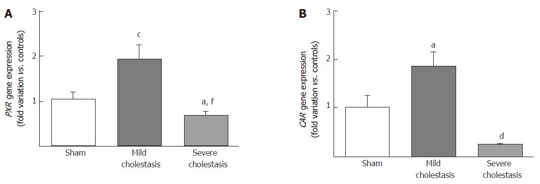
Figure 5 mRNA expression of nuclear receptors in rat livers.
Pregnane x receptor (A) and constitutive androstane receptor (B) gene expression in sham-operated and cholestatic rats reported as fold variations compared with sham rats. Results are mean ± SEM obtained from 8 rats in each group. aP < 0.05, cP < 0.001 vs sham rats; dP < 0.05, fP < 0.01 vs mildly cholestatic rats.
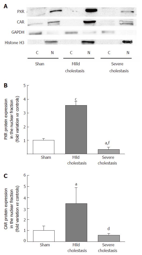
Figure 6 Nuclear protein expression of nuclear receptors in rat livers.
Representative Western blot membrane showing pregnane x receptor and constitutive androstane receptor protein expression in the cytoplasmic and nuclear fractions of sham and cholestatic rats (A). GAPDH and Histone H3 were used to assess the purity of the cytoplasmic and nuclear fractions, respectively. Densitometric analysis of proteins in the nuclear fraction representing the PXR (B) and CAR (C) nuclear expression. Results are expressed as mean ± SEM obtained from 8 rats in each group. aP < 0.05, cP < 0.001 vs sham rats; dP < 0.05, fP < 0.001 vs mildly cholestatic rats.
- Citation: Gabbia D, Pozza AD, Albertoni L, Lazzari R, Zigiotto G, Carrara M, Baldo V, Baldovin T, Floreani A, Martin SD. Pregnane X receptor and constitutive androstane receptor modulate differently CYP3A-mediated metabolism in early- and late-stage cholestasis. World J Gastroenterol 2017; 23(42): 7519-7530
- URL: https://www.wjgnet.com/1007-9327/full/v23/i42/7519.htm
- DOI: https://dx.doi.org/10.3748/wjg.v23.i42.7519


