Copyright
©The Author(s) 2017.
World J Gastroenterol. Nov 14, 2017; 23(42): 7505-7518
Published online Nov 14, 2017. doi: 10.3748/wjg.v23.i42.7505
Published online Nov 14, 2017. doi: 10.3748/wjg.v23.i42.7505
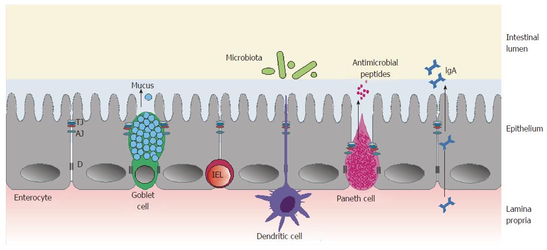
Figure 1 Schematic illustration of the intestinal barrier.
The three main components of the intestinal barrier: the microbiota; epithelium, with its specialized cells (goblet cells, Paneth cells and enterocytes), together with a layer of mucus; and gut-associated lymphoid tissue cells, including IELs and dendritic cells. AJ: Adherens junction; D: Desmosome; IEL: Intraepithelial lymphocyte; TJ: Tight junction.
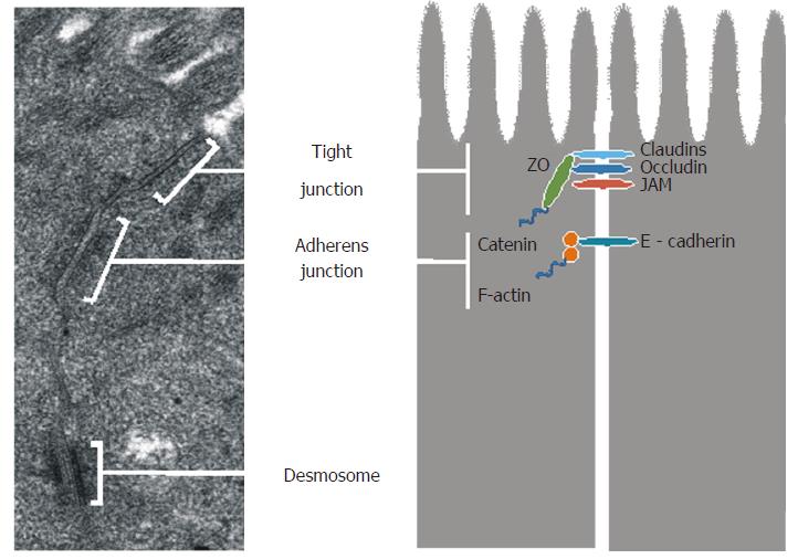
Figure 2 Ultrastructure and corresponding schematic representation of intercellular junctions.
Transmission electron microscopy (JEOL JEM-1011, Japan; × 60000) was used to show the ultrastructure of intercellular junctions in the human small intestine. The transmission electron micrograph comes from our own research. JAM: Junction adhesion molecule.
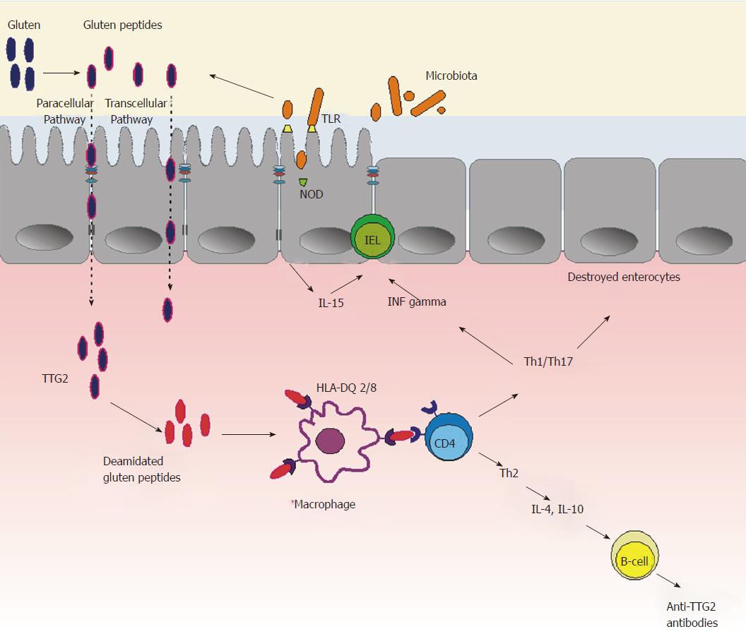
Figure 3 Schematic illustration of celiac disease pathogenesis.
Microbiota dysbiosis activates innate immunity resulting in pro-inflammatory changes, which leads to IEL infiltration and epithelial barrier disruption. This ultimately results in an increased paracellular and transcellular transfer of immunogenic gluten peptides and activation of adaptive pro-inflammatory Th1/Th17 pathways, leading to villous atrophy and production of autoantibodies against intestinal TTG2. HLA: Human leucocyte antigen; IEL: Intraepithelial lymphocyte; IL: Interleukin; INF: Interferon; NOD: Nucleotide-binding oligomerization domain; Th: T helper; TLR: Toll-like receptor; TTG2: Tissue transglutaminase 2.
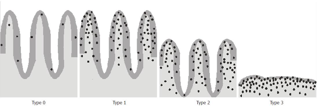
Figure 4 A schematic illustration of progressive histopathological changes in the small intestine according to the modified Marsh-Oberhuber grading scale.
Type 0: Normal mucosa with IEL count < 25 per 100 enterocytes; Type 1: Normal mucosa with an increased IEL count; Types 2 and 3 show increased IEL counts and lymphocytes in the lamina propria. IELs are presented as black dots. IEL: Intraepithelial lymphocyte.
- Citation: Cukrowska B, Sowińska A, Bierła JB, Czarnowska E, Rybak A, Grzybowska-Chlebowczyk U. Intestinal epithelium, intraepithelial lymphocytes and the gut microbiota - Key players in the pathogenesis of celiac disease. World J Gastroenterol 2017; 23(42): 7505-7518
- URL: https://www.wjgnet.com/1007-9327/full/v23/i42/7505.htm
- DOI: https://dx.doi.org/10.3748/wjg.v23.i42.7505


