Copyright
©The Author(s) 2017.
World J Gastroenterol. Nov 7, 2017; 23(41): 7489-7494
Published online Nov 7, 2017. doi: 10.3748/wjg.v23.i41.7489
Published online Nov 7, 2017. doi: 10.3748/wjg.v23.i41.7489
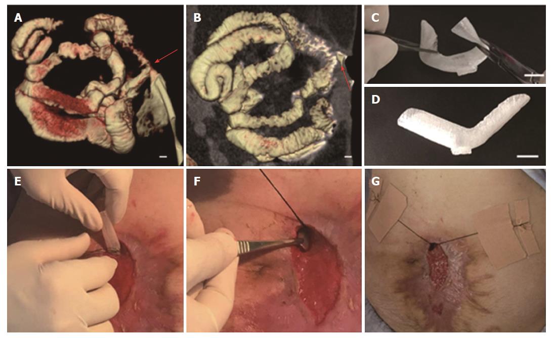
Figure 1 Fabrication and implantation of 3D-printed fistula stent.
A and B: 3D-reconstructed fistulography of the ECF: the diameters of the proximal and distal GI tract were 15 mm and 12 mm, respectively, bar = 1 mm (red arrow: ECF); C: Flexibility of fistula stent made of TPU, bar = 15 mm; D: Shape memory of the fistula stent after application of an external force, bar = 15 mm; E and G: The process of the fistula stent implantation. ECF: Enterocutaneous fistula.
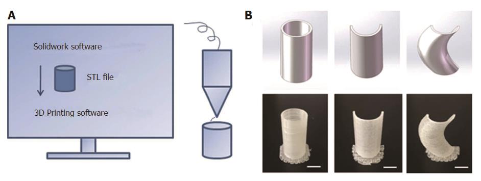
Figure 2 3D printing technique.
A: Schematic diagram of the 3D printing technique; B: 3D-printed models: Column, semi-column and tortuous semi-column, bar = 1 cm.
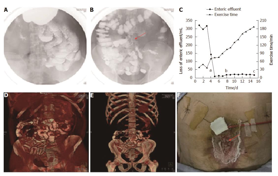
Figure 3 Follow-up after stent implantation.
A: Image of small intestine after barium meal, indicating no obstruction; B: Image of colon after barium meal, indicating that contrast passed through the stent smoothly (red arrow: fistula stent); C: Loss of enteric effluent and the time of exercise tolerance before and after stent implantation. aTime point of stent insertion; bTime point of starting EN; cTime point of skin transplantation; D and E: 3D-reconstructed images after barium meal, indicating no abnormity after starting EN; G: Skin transplantation for abdominal wall reconstruction (red arrow: skin graft). EN: Enteral nutrition.
- Citation: Huang JJ, Ren JA, Wang GF, Li ZA, Wu XW, Ren HJ, Liu S. 3D-printed “fistula stent” designed for management of enterocutaneous fistula: An advanced strategy. World J Gastroenterol 2017; 23(41): 7489-7494
- URL: https://www.wjgnet.com/1007-9327/full/v23/i41/7489.htm
- DOI: https://dx.doi.org/10.3748/wjg.v23.i41.7489


