Copyright
©The Author(s) 2017.
World J Gastroenterol. Oct 14, 2017; 23(38): 6952-6961
Published online Oct 14, 2017. doi: 10.3748/wjg.v23.i38.6952
Published online Oct 14, 2017. doi: 10.3748/wjg.v23.i38.6952
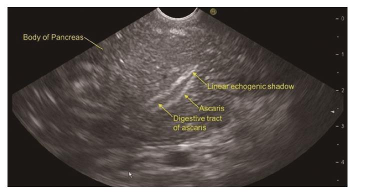
Figure 1 Linear endoscopic ultrasound from the stomach showing Ascaris lumbricoides in the body of the pancreas.
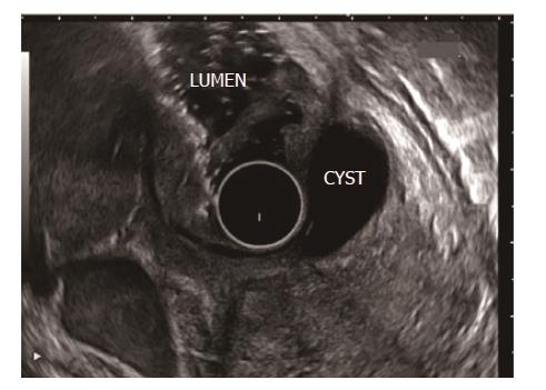
Figure 2 Radial endoscopic ultrasound from the duodenum shows duodenal duplication cyst as etiology of idiopathic pancreatitis.
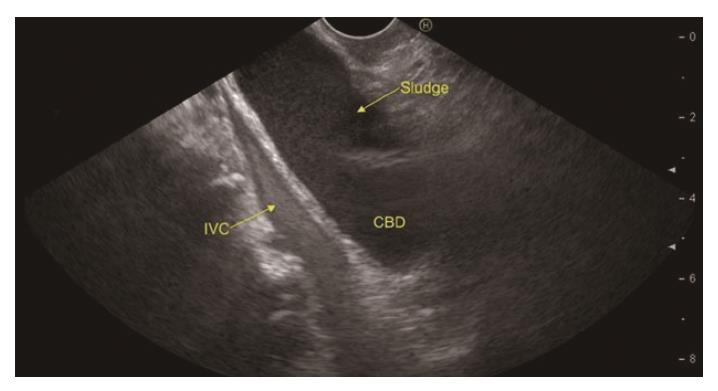
Figure 3 Linear endoscopic ultrasound from the duodenal bulb shows echogenic biliary sludge in the common bile duct.
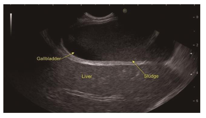
Figure 4 Linear endoscopic ultrasound from the duodenal bulb shows echogenic sludge in the gallbladder.
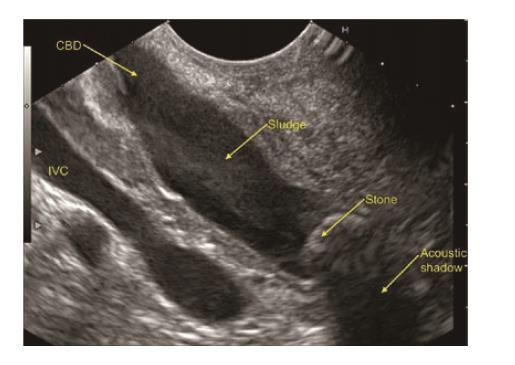
Figure 5 Common bile duct stone with sludge seen on linear endoscopic ultrasound from the duodenal bulb in a 36-year-old male presenting with 3 episodes of idiopathic acute pancreatitis in last 7 mo.
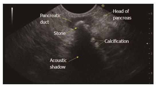
Figure 6 Features of chronic pancreatitis seen on linear endoscopic ultrasound from descending duodenum.
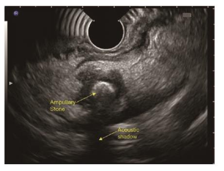
Figure 7 Ampullary stone with acoustic shadow seen on radial endoscopic ultrasound from descending duodenum.
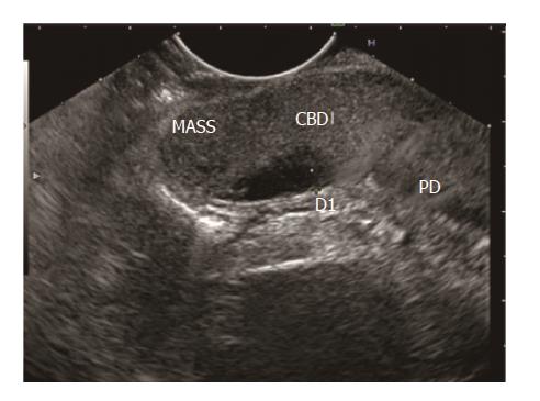
Figure 8 Hypoechoic ampullary mass extending into common bile duct.
- Citation: Somani P, Sunkara T, Sharma M. Role of endoscopic ultrasound in idiopathic pancreatitis. World J Gastroenterol 2017; 23(38): 6952-6961
- URL: https://www.wjgnet.com/1007-9327/full/v23/i38/6952.htm
- DOI: https://dx.doi.org/10.3748/wjg.v23.i38.6952


