Copyright
©The Author(s) 2017.
World J Gastroenterol. Sep 21, 2017; 23(35): 6491-6499
Published online Sep 21, 2017. doi: 10.3748/wjg.v23.i35.6491
Published online Sep 21, 2017. doi: 10.3748/wjg.v23.i35.6491
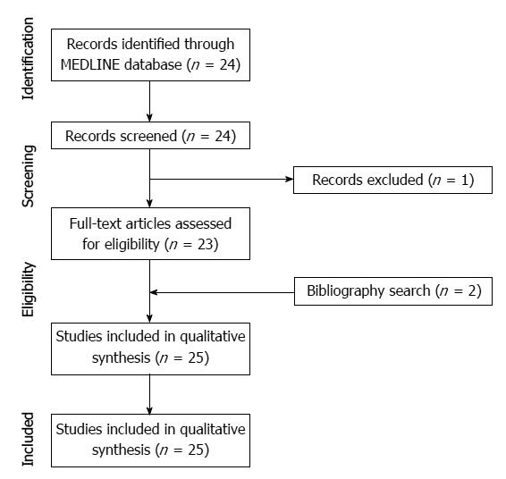
Figure 1 Flow diagram for the selection of studies.
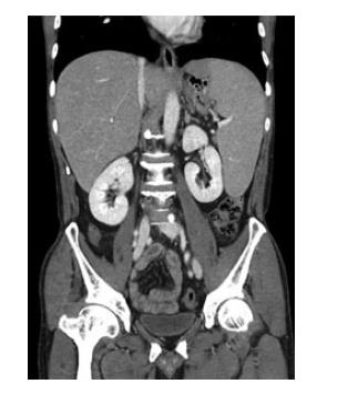
Figure 2 On a coronal computed tomography image taken 2 mo after autologous stem-cell transplantation, the spleen was enlarged, measuring 17 cm in the longest dimension, and indicative of recurred lymphoma.
The enlarged spleen abutted to the gastric fundus.
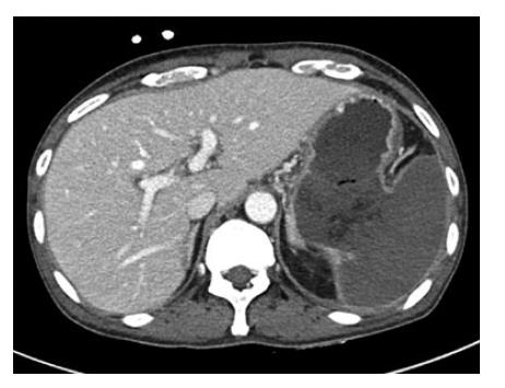
Figure 3 On an axial computed tomography image taken after chemotherapy, a huge fistula was shown between the gastric lumen and the spleen.
The spleen was totally infarcted.
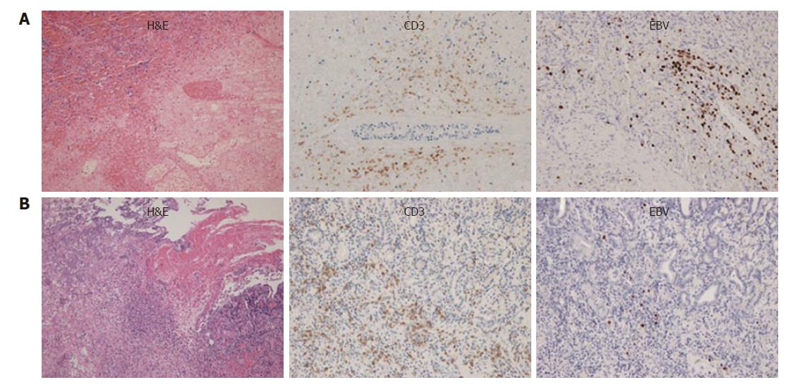
Figure 4 Microscopic specimen of the spleen and stomach.
A: Atypical lymphoma cells were found in the spleen on hematoxylin-eosin stain (left). These cells showed positivity for CD3 on immunohistochemistry stain (middle) and EBV on EBV RNA stain (right). There was also extensive coagulative necrosis indicative of splenic infarction; B: Lymphoma cells were found in the stomach wall near the gastrosplenic fistula on hematoxylin-eosin stain (left), and which also showed positivity for CD3 (middle) and EBV (right). NK/T-cell lymphoma was diagnosed. These findings suggested that lymphoma cells may have infiltrated from the spleen to the stomach wall through the perforation site. EBV: Epstein-Barr virus.
- Citation: Kang DH, Huh J, Lee JH, Jeong YK, Cha HJ. Gastrosplenic fistula occurring in lymphoma patients: Systematic review with a new case of extranodal NK/T-cell lymphoma. World J Gastroenterol 2017; 23(35): 6491-6499
- URL: https://www.wjgnet.com/1007-9327/full/v23/i35/6491.htm
- DOI: https://dx.doi.org/10.3748/wjg.v23.i35.6491


