Copyright
©The Author(s) 2017.
World J Gastroenterol. Sep 21, 2017; 23(35): 6457-6466
Published online Sep 21, 2017. doi: 10.3748/wjg.v23.i35.6457
Published online Sep 21, 2017. doi: 10.3748/wjg.v23.i35.6457
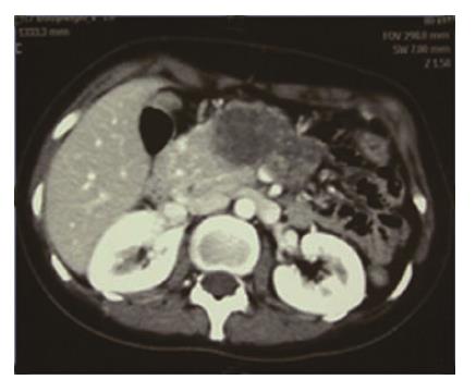
Figure 1 Preoperative computed tomography view of a 26-year-old female patient with a diagnosis of pseudo-papillary tumors.
The figure shows that the lesion occupied the body and neck region with necrosis of the tail on preoperative computed tomography imaging. Normal pancreatic tissue is observed in the ventral pancreas region.
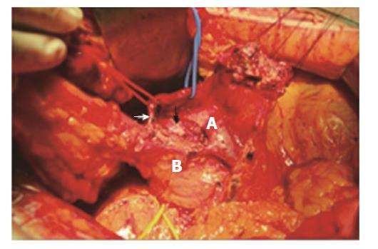
Figure 2 Intraoperative view of duodenum and ventral pancreas preserving subtotal pancreatectomy.
A: Superior mesenteric vein; B: Ventral pancreas. Black arrow: intrapancreatic bile duct; White arrow: gastroduodenal artery.
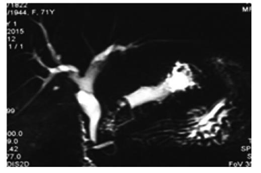
Figure 3 Preoperative magnetic resonance imaging view of a 71-year-old female patient with diagnosis of MD-IPMN.
The figure shows that the lesion involves the whole dorsal duct (including Santorini duct). The diameter of ventral pancreatic duct is normal on the MRCP image.
- Citation: Wang X, Tan CL, Song HY, Yao Q, Liu XB. Duodenum and ventral pancreas preserving subtotal pancreatectomy for low-grade malignant neoplasms of the pancreas: An alternative procedure to total pancreatectomy for low-grade pancreatic neoplasms. World J Gastroenterol 2017; 23(35): 6457-6466
- URL: https://www.wjgnet.com/1007-9327/full/v23/i35/6457.htm
- DOI: https://dx.doi.org/10.3748/wjg.v23.i35.6457


