Copyright
©The Author(s) 2017.
World J Gastroenterol. Sep 21, 2017; 23(35): 6448-6456
Published online Sep 21, 2017. doi: 10.3748/wjg.v23.i35.6448
Published online Sep 21, 2017. doi: 10.3748/wjg.v23.i35.6448
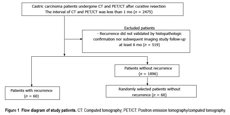
Figure 1 Flow diagram of study patients.
CT: Computed tomography; PET/CT: Positron emission tomography/computed tomography.
Figure 2 Peritoneal carcinomatosis detected only on contrast-enhanced computed tomography in a 46-year-old woman who underwent gastrectomy for advanced gastric cancer 17 mo prior.
A: Contrast-enhanced abdominal computed tomography shows enhanced peritoneal thickening (arrow) with ascites. Peritoneal metastasis was confirmed by histological analysis of ascitic fluid. B: Positron emission tomography/computed tomography image at the same level without evident hypermetabolism in the peritoneum.
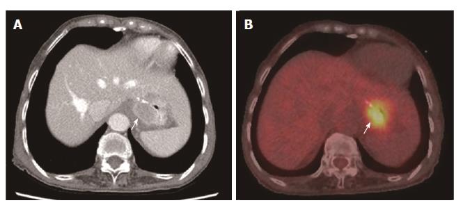
Figure 3 Locoregional recurrence detected on both contrast-enhanced computed tomography and positron emission tomography/computed tomography in a 76-year-old woman who underwent gastrectomy for advanced gastric cancer 21 mo prior.
A: Contrast-enhanced abdominal computed tomography shows enhanced wall thickening (arrow) at the anastomotic site; B: Positron emission tomography/computed tomography image at the same level shows hypermetabolism at the anastomotic site.
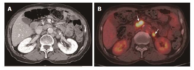
Figure 4 Lymph node recurrence detected on both contrast-enhanced computed tomography and positron emission tomography/computed tomography in a 73-year-old man who underwent gastrectomy 18 mo prior.
A: Contrast-enhanced abdominal computed tomography shows enlarged lymph nodes at the aortocaval and para-aortic spaces; B: Positron emission tomography/computed tomography image at the same level shows hypermetabolism at the aortocaval and para-aortic spaces.
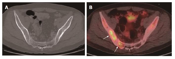
Figure 5 Bone metastasis detected only on positron emission tomography/computed tomography in a 47-year-old man who underwent gastrectomy for advanced gastric cancer 16 mo prior.
A: Contrast-enhanced abdominal computed tomography showing the absence of bone lesions; B: Positron emission tomography/computed tomography image at the same level showing multiple hypermetabolic lesions in the pelvic bones and sacrum.
- Citation: Kim JH, Heo SH, Kim JW, Shin SS, Min JJ, Kwon SY, Jeong YY, Kang HK. Evaluation of recurrence in gastric carcinoma: Comparison of contrast-enhanced computed tomography and positron emission tomography/computed tomography. World J Gastroenterol 2017; 23(35): 6448-6456
- URL: https://www.wjgnet.com/1007-9327/full/v23/i35/6448.htm
- DOI: https://dx.doi.org/10.3748/wjg.v23.i35.6448


