Copyright
©The Author(s) 2017.
World J Gastroenterol. Sep 14, 2017; 23(34): 6252-6260
Published online Sep 14, 2017. doi: 10.3748/wjg.v23.i34.6252
Published online Sep 14, 2017. doi: 10.3748/wjg.v23.i34.6252
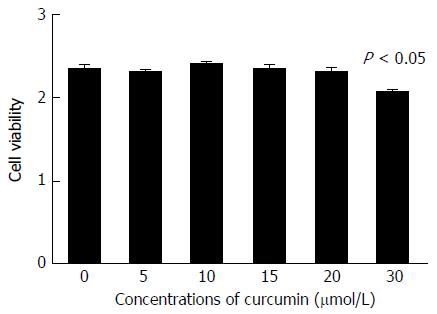
Figure 1 Cell cytotoxicity of curcumin.
HepG2.2.15 cells were treated with 0, 5, 10, 15, 20 or 30 μmol/L curcumin for 2 d and then subjected to CCK-8 assay to detect toxic effect. The experiment was performed in duplicate and repeated at least three times.
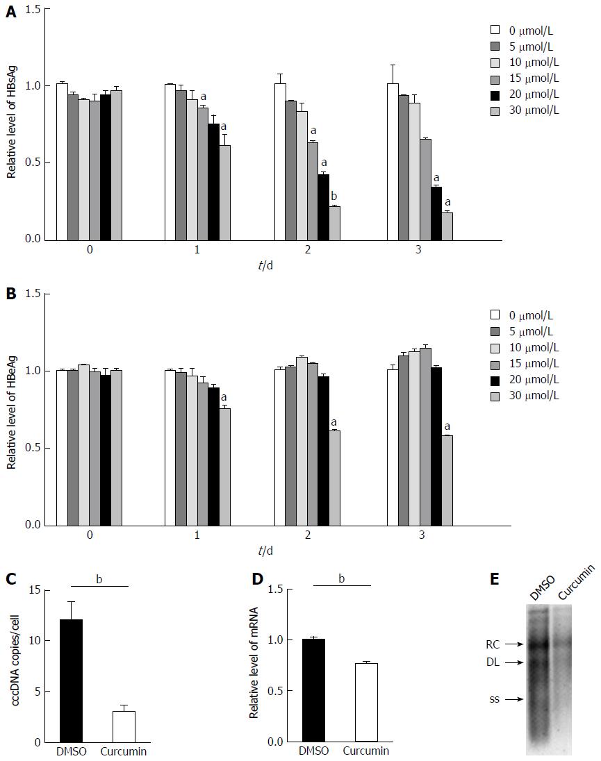
Figure 2 Curcumin inhibits hepatitis B virus replication and expression.
A: HepG2.215 cells were treated with 0, 5, 10, 15, 20 or 30 μmol/L curcumin for three consecutive days. Culture medium from each day was collected and analysed for levels of HBsAg; B: Culture medium from each day was collected and analysed for levels of HBeAg; C: Hepatitis B virus (HBV) cccDNA accumulation in HepG2.215 cells treated with 20 μmol/L curcumin or DMSO for 2 d. HBV cccDNA was digested with Plasmid-Safe ATP-Dependent DNase to degrade contaminating HBV that had inserted in cellular genomic DNA and OC species and was then subjected to PCR amplification to amplify HBV cccDNA forms. Results are expressed as numbers of cccDNA copies per cell; D: Total RNA was extracted from HepG2.215 cells treated with 20 μmol/L curcumin or DMSO for 2 d and was subjected to real-time PCR to quantify HBV mRNA transcript levels; E: HBV DNA was extracted from intracellular core particles in HepG2.215 cells treated with 20 μmol/L curcumin or DMSO for 2 d. Southern blot analysis of HBV DNA replicative intermediates. RC, DL and SS represent relaxed circular, double linear and single-stranded forms of HBV DNA, respectively. All experiments were repeated at least three times; ELISA and RT-PCR were performed in duplicate. aP < 0.05; bP < 0.01. cccDNA: Covalently closed circular DNA; DMSO: Dimethyl sulphoxide.
Figure 3 Curcumin mediates chromosomal and covalently closed circular DNA-bound histone deacetylation.
A: HepG2.215 cells were treated with 0, 1, 2.5, 5 or 10 μmol/L curcumin and incubated at 37 °C for 2 d. The acetylation status of cellular H3 histones was analysed by Western blot; B: HepG2.215 cells were treated with 20 μmol/L curcumin for the indicated periods of time; C: ChIP on HepG2.215 cells treated with 20 μmol/L curcumin or DMSO for 2 d was performed using specific antibodies to acetyl-histone H3 (AcH3), acetyl-histone H4 (AcH4) or control IgG. Immunoprecipitated chromatins were digested with Plasmid-Safe ATP-Dependent DNase to degrade contaminating HBV that had inserted in cellular genomic DNA and OC species and then were subjected to PCR amplification to select HBV cccDNA forms. All experiments were repeated at least three times. cccDNA: Covalently closed circular DNA; HBV: Hepatitis B virus; DMSO: Dimethyl sulphoxide.
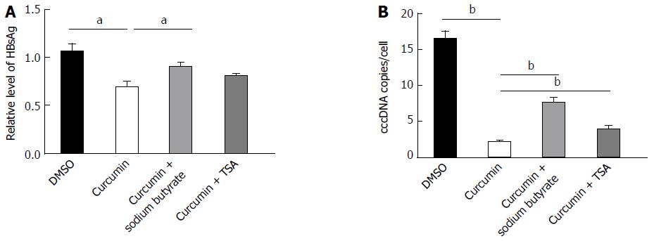
Figure 4 Histone deacetylase inhibitors block the inhibitory effect of curcumin.
A: HepG2.215 cells were treated with 20 μmol/L curcumin alone or with either 5 mmol/L sodium butyrate or 1 μmol/L TSA for 2 d. Medium was collected and analysed for levels of HBsAg; B: HBV cccDNA was extracted and subjected to real-time qPCR. All experiments were performed in duplicate and repeated at least three times. aP < 0.05; bP < 0.01. cccDNA: Covalently closed circular DNA; DMSO: Dimethyl sulphoxide; TSA: Trichostatin A; HBV: Hepatitis B virus; HBsAg: HBV surface antigen.
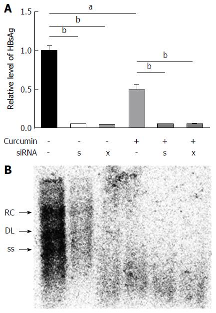
Figure 5 siRNAs against hepatitis B virus enhance the inhibitory effects of curcumin.
HepG2.215 cells were transfected with 20 nmol/L siRNAs or negative control (HC) siRNA and were treated with 20 μmol/L curcumin or dimethyl sulphoxide for the next 2 d. HBsAg in cell culture supernatants and intracellular HBV replicative intermediates were detected by ELISA (A) and Southern blot analysis (B), respectively. All experiments were repeated at least three times; ELISA was performed in duplicate. aP < 0.05; bP < 0.01. HBV: Hepatitis B virus; HBsAg: HBV surface antigen.
- Citation: Wei ZQ, Zhang YH, Ke CZ, Chen HX, Ren P, He YL, Hu P, Ma DQ, Luo J, Meng ZJ. Curcumin inhibits hepatitis B virus infection by down-regulating cccDNA-bound histone acetylation. World J Gastroenterol 2017; 23(34): 6252-6260
- URL: https://www.wjgnet.com/1007-9327/full/v23/i34/6252.htm
- DOI: https://dx.doi.org/10.3748/wjg.v23.i34.6252


