Copyright
©The Author(s) 2017.
World J Gastroenterol. Sep 14, 2017; 23(34): 6201-6211
Published online Sep 14, 2017. doi: 10.3748/wjg.v23.i34.6201
Published online Sep 14, 2017. doi: 10.3748/wjg.v23.i34.6201
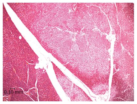
Figure 1 Light microscopy image of the porcine pancreas after double-balloon enteroscopy showing located ischemic necrosis in pancreatic interlobular tissue.
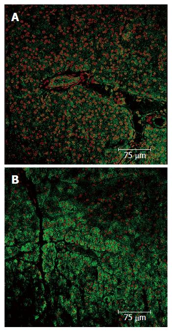
Figure 2 Pancreas immunohistochemistry.
A: Expression of the VEGF in a normal parenchyma; B: Pancreatic acini with normal structure and nuclei, which express VEGF. In upper right corner a ischemic zone where less VEGF expression is shown, the pancreatic acini structure has been lost and nuclei are pyknotic. VEGF: Vascular endothelial growth factor.
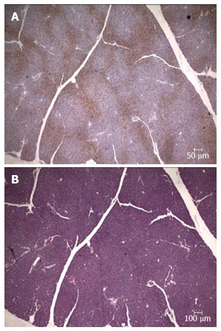
Figure 3 Pancreas immunohistochemistry.
A: Expression of the pimonidazole hydrochloride (Hypoxyprobe®) presence of focal areas positively stained; B: Serial section stained with hematoxyline-eosine.
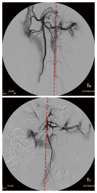
Figure 4 Selective angiogram of the cranial mesenteric artery.
T0: Before the DBE; T1: With the endoscope inserted at maximum during DBE. Red line shows the aorta topography. DBE: Double balloon enteroscopy.
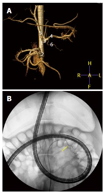
Figure 5 Computed tomography angiogram (A) with vascular 3D reconstruction to monitor his individual vascular anatomy (1) abdominal aorta; (2) cranial mesenteric artery; (3) celiac artery; (4) splenic artery; (5) hepatic artery; and (6) major pancreatic artery.
B: Super-selective angiography of the major pancreatic artery (arrow) with the endoscope inserted at maximum during DBE. The nuclear dye Hoechst (Bizbenzimida H 33258 fluorochrome) is injected through that vessel to allow its contact with the cells in the tail of the pancreas. DBE: Double balloon enteroscopy.
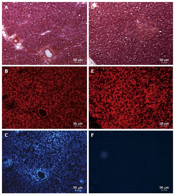
Figure 6 Fluorescence microscopy image of cryosections from left lobe of the pancreas showing distribution of endocrine and exocrine cells with nuclei stained in disto-medial (A-C) and proximo-lateral (D-F) portions.
A and D: Serial sections stained with hematoxyline-eosine; B-E: Serial sections stained with the nuclear marker TO-PRO®3 iodide; C and F: Serial sections showing the location the nuclear dye Hoechst (Bizbenzimida H 33258 fluorochrome).
- Citation: Latorre R, López-Albors O, Soria F, Morcillo E, Esteban P, Pérez-Cuadrado-Robles E, Pérez-Cuadrado-Martínez E. Evidences supporting the vascular etiology of post-double balloon enteroscopy pancreatitis: Study in porcine model. World J Gastroenterol 2017; 23(34): 6201-6211
- URL: https://www.wjgnet.com/1007-9327/full/v23/i34/6201.htm
- DOI: https://dx.doi.org/10.3748/wjg.v23.i34.6201


